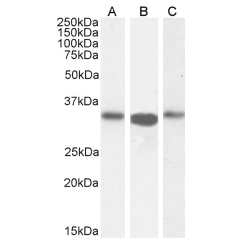
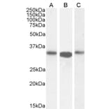
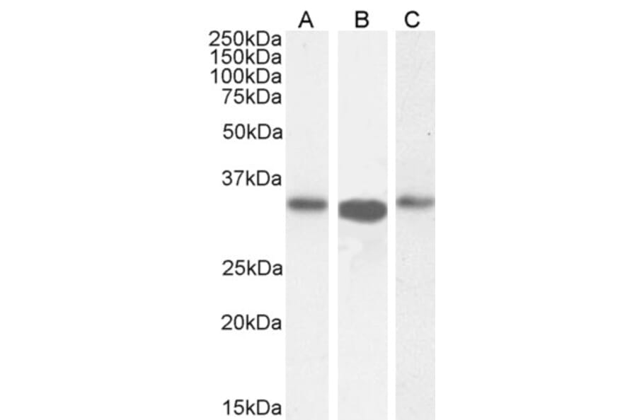
ARPC2 expression in Caco-2 (A), HeLa (B), and Jurkat (C) lysates analyzed by western blot. Cells were lysed in RIPA buffer and 35µg protein was run per lane. Primary incubation was performed with Anti-ARPC2 Antibody (A83514) at 0.5µg/ml (A) or 0.1µg/ml (B-C) and detected by chemiluminescence.
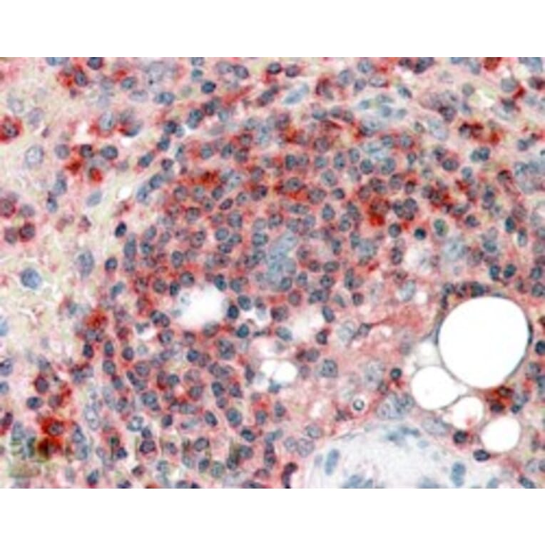
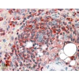
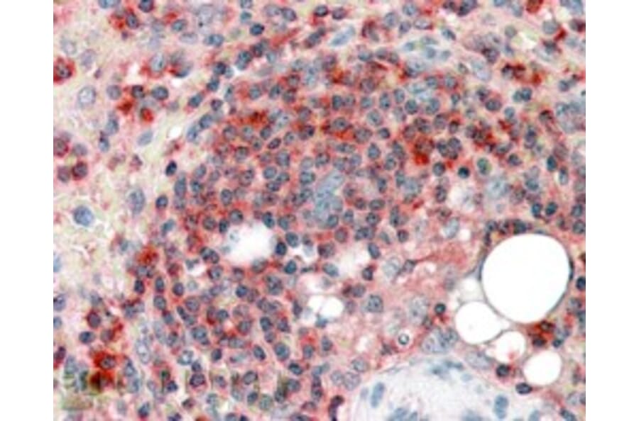
ARPC2 expression in Human Spleen analyzed by immunohistochemistry. Tissue was paraffin-embedded, and antigen retrieval was achieved by steaming in citrate buffer, pH 6. Staining was performed with Anti-ARPC2 Antibody (A83514) at 3.8µg/ml and revealed with alkaline phosphatase (AP).
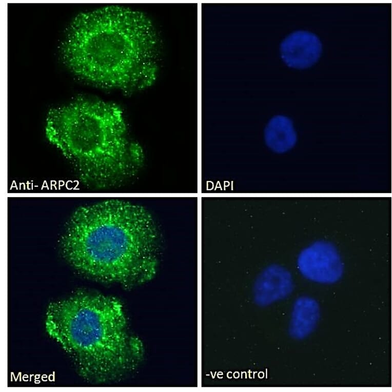
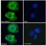
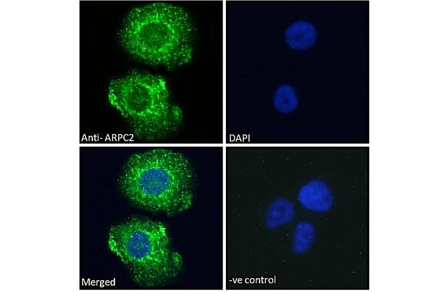
ARPC2 expression in A431 cells analyzed by immunofluorescence. Cells were permeabilized with 0.15% Triton. Staining was performed with Anti-ARPC2 Antibody (A83514) at 10µg/ml for 1 hour and Alexa Fluor 488 secondary antibody at 2µg/ml. Cytoplasmic staining shown and nuclei were stained with DAPI (blue). Negative control: Goat IgG Isotype Control at 10µg/ml followed by Alexa Fluor 488 secondary antibody at 2µg/ml.
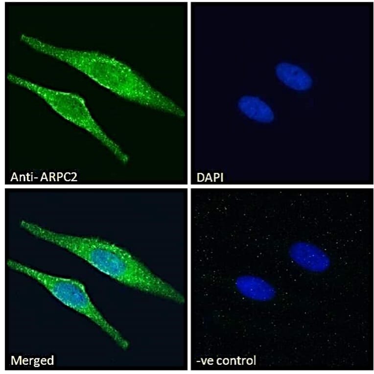
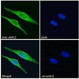
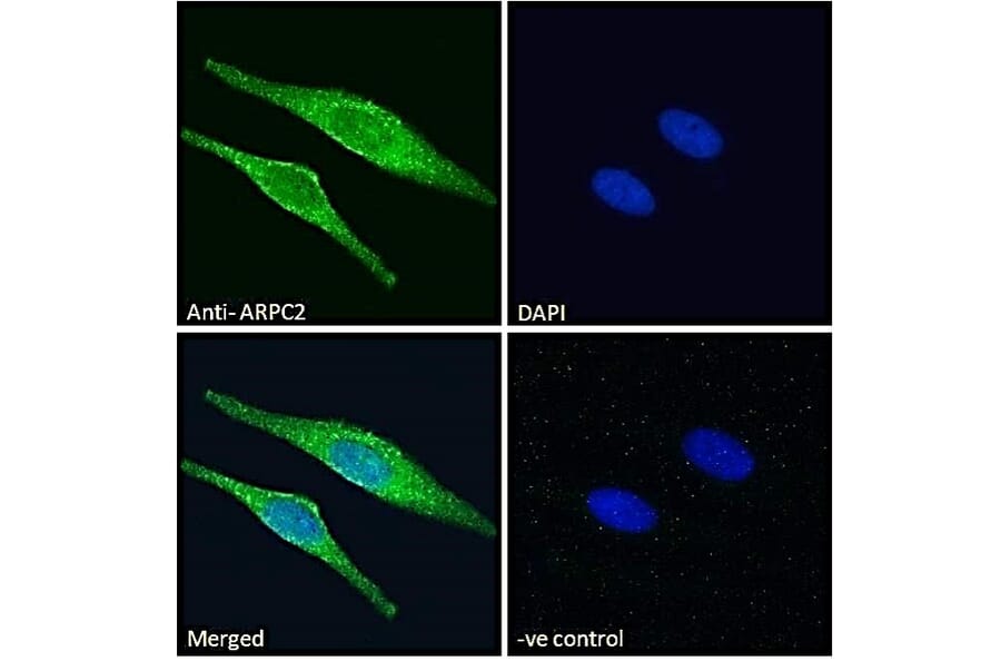
ARPC2 expression in HeLa cells analyzed by immunofluorescence. Cells were permeabilized with 0.15% Triton. Staining was performed with Anti-ARPC2 Antibody (A83514) at 10µg/ml for 1 hour and Alexa Fluor 488 secondary antibody at 2µg/ml. Cytoplasmic staining shown and nuclei were stained with DAPI (blue). Negative control: Goat IgG Isotype Control at 10µg/ml followed by Alexa Fluor 488 secondary antibody at 2µg/ml.
Publishing research using Anti-ARPC2 Antibody (A83514)? Please let us know so that we can list the citation on this page.
Alternative products to Anti-ARPC2 Antibody (A83514)


![Western Blot - Anti-ARPC2 Antibody [ARC2558] (A305917) - Antibodies.com](https://cdn.antibodies.com/image/catalog/305/A305917_1.jpg?profile=product_alternative)