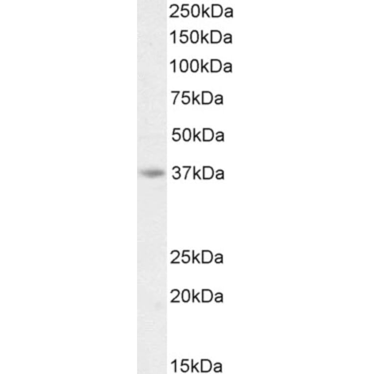
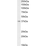
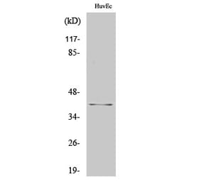
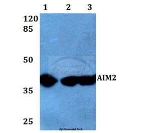
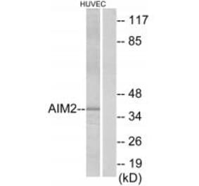
AIM2 expression in HeLa cell lysate analyzed by western blot. Cells were lysed in RIPA buffer and 35µg protein was run per lane. Primary antibody incubation was performed with Anti-AIM2 Antibody (A84784) at 0.75µg/ml and detected by chemiluminescence.
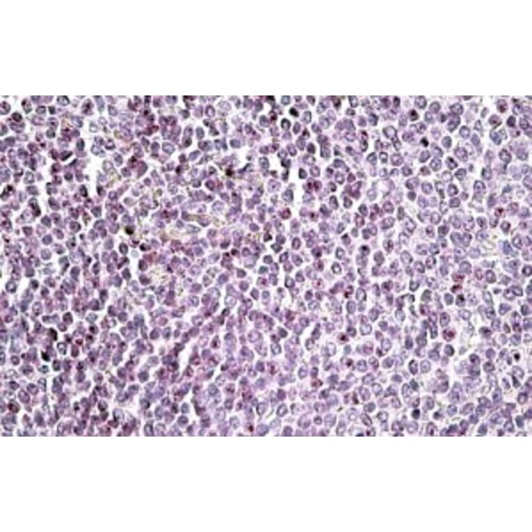
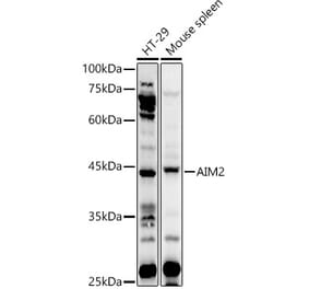
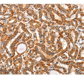
AIM2 expression in Human Spleen analyzed by immunohistochemistry. Tissue was paraffin-embedded, and antigen retrieval was achieved by steaming in citrate buffer, pH 6. Staining was performed with Anti-AIM2 Antibody (A84784) at 3.75µg/ml and revealed with alkaline phosphatase (AP).
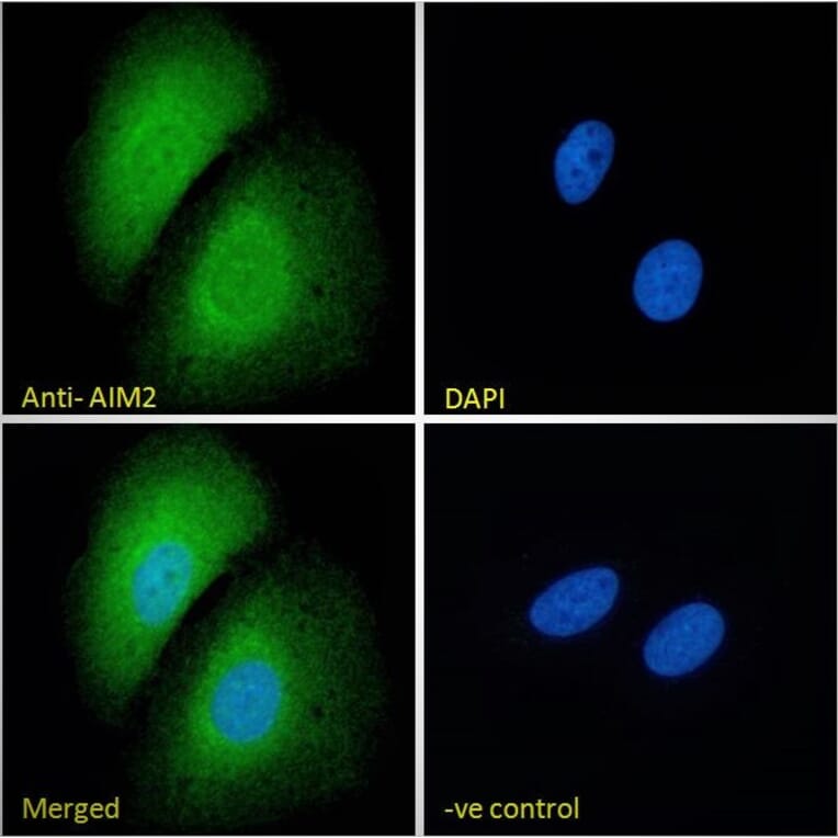
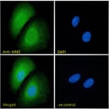
AIM2 expression in U2OS cells analyzed by immunofluorescence. Cells were permeabilized with 0.15% Triton. Staining was performed with Anti-AIM2 Antibody (A84784) at 10µg/ml for 1 hour and Alexa Fluor 488 secondary antibody at 2µg/ml. Cytoplasmic and some nuclear staining shown and nuclei were stained with DAPI (blue). Negative control: Goat IgG Isotype Control at 10µg/ml followed by Alexa Fluor 488 secondary antibody at 2µg/ml.
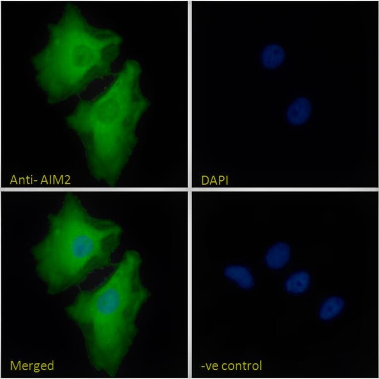
AIM2 expression in HeLa cells analyzed by immunofluorescence. Cells were permeabilized with 0.15% Triton. Staining was performed with Anti-AIM2 Antibody (A84784) at 10µg/ml for 1 hour and Alexa Fluor 488 secondary antibody at 2µg/ml. Cytoplasmic and some nuclear staining shown and nuclei were stained with DAPI (blue). Negative control: Goat IgG Isotype Control at 10µg/ml followed by Alexa Fluor 488 secondary antibody at 2µg/ml.
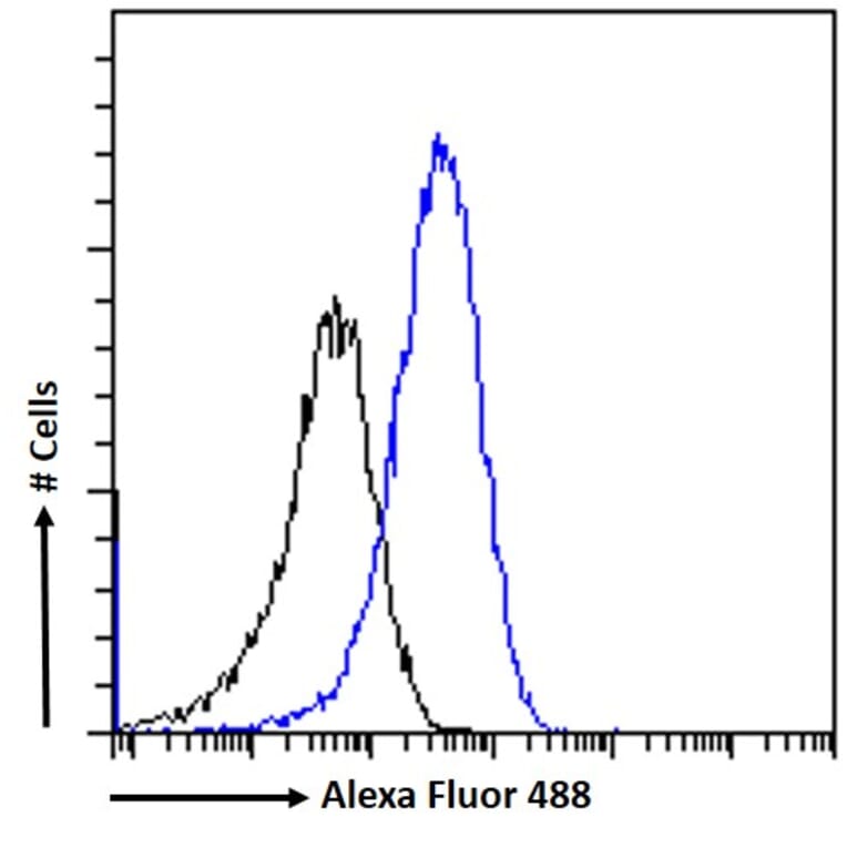
AIM2 expression in HeLa cells (blue line) analyzed by flow cytometry. Cells were fixed in PFA and permeabilized with 0.5% Triton. Staining was performed with Anti-AIM2 Antibody (A84784) at 10µg/ml for 1 hour and Alexa Fluor 488 secondary antibody at 1µg/ml. Negative Control: Goat IgG Isotype Control (black line) followed by Alexa Fluor 488 secondary antibody.