Mouse monoclonal [LH7.2] antibody to Collagen VII.
Specificity
This antibody recognizes an epitope located on collagenase digested Type VII collagen (150kDa), i.e. the non-helical carboxyl terminal region of the dimer. Binding is localized at the inferior border of the lamina densa as demonstrated by immunoelectron microscopy. Type VII is also found in the retina, where its function is unknown. It interacts with Laminin 5 and Fibronectin. Collagen VII is a stratified squamous epithelial basement membrane protein that forms anchoring fibrils which may contribute to epithelial basement membrane organization and adherence by interacting with extracellular matrix (ECM) proteins such as type IV collagen.
SDS-PAGE - Anti-Collagen VII Antibody [LH7.2] (A248248)
SDS-PAGE analysis of Anti-Collagen VII Antibody [LH7.2] under non-reduced and reduced conditions; showing intact IgG and intact heavy and light chains, respectively. SDS-PAGE analysis confirms the integrity and purity of the antibody.
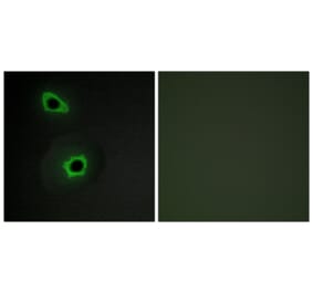
Immunofluorescence - Anti-Collagen VII Antibody [LH7.2] (A248248)
Immunofluorescent analysis of PFA fixed HeLa cells stained with Anti-Collagen VII Antibody [LH7.2] followed by Goat Anti-Mouse IgG (CF® 488) (Green). The nuclear counterstain is RedDot.
Flow Cytometry - Anti-Collagen VII Antibody [LH7.2] (A248248)
Flow cytometric analysis of PFA fixed HeLa cells using Anti-Collagen VII Antibody [LH7.2] followed by Goat Anti-Mouse IgG (CF® 488) (Blue). Isotype Control (Red).


![Immunohistochemistry - Anti-Collagen VII Antibody [LH7.2] (A248248) - Antibodies.com](https://cdn.antibodies.com/image/catalog/248/A248248_1.jpg?profile=product_top)
![Immunohistochemistry - Anti-Collagen VII Antibody [LH7.2] (A248248) - Antibodies.com](https://cdn.antibodies.com/image/catalog/248/A248248_2.jpg?profile=product_top)
![SDS-PAGE - Anti-Collagen VII Antibody [LH7.2] (A248248) - Antibodies.com](https://cdn.antibodies.com/image/catalog/248/A248248_3.jpg?profile=product_top)
![Immunofluorescence - Anti-Collagen VII Antibody [LH7.2] (A248248) - Antibodies.com](https://cdn.antibodies.com/image/catalog/248/A248248_4.jpg?profile=product_top)
![Flow Cytometry - Anti-Collagen VII Antibody [LH7.2] (A248247) - Antibodies.com](https://cdn.antibodies.com/image/catalog/248/A248248_5.jpg?profile=product_top)
![Immunohistochemistry - Anti-Collagen VII Antibody [LH7.2] (A248248) - Antibodies.com](https://cdn.antibodies.com/image/catalog/248/A248248_1.jpg?profile=product_top_thumb)
![Immunohistochemistry - Anti-Collagen VII Antibody [LH7.2] (A248248) - Antibodies.com](https://cdn.antibodies.com/image/catalog/248/A248248_2.jpg?profile=product_top_thumb)
![SDS-PAGE - Anti-Collagen VII Antibody [LH7.2] (A248248) - Antibodies.com](https://cdn.antibodies.com/image/catalog/248/A248248_3.jpg?profile=product_top_thumb)
![Immunofluorescence - Anti-Collagen VII Antibody [LH7.2] (A248248) - Antibodies.com](https://cdn.antibodies.com/image/catalog/248/A248248_4.jpg?profile=product_top_thumb)
![Flow Cytometry - Anti-Collagen VII Antibody [LH7.2] (A248247) - Antibodies.com](https://cdn.antibodies.com/image/catalog/248/A248248_5.jpg?profile=product_top_thumb)
![Immunohistochemistry - Anti-Collagen VII Antibody [LH7.2] (A248248) - Antibodies.com](https://cdn.antibodies.com/image/catalog/248/A248248_1.jpg?profile=product_image)
![Immunohistochemistry - Anti-Collagen VII Antibody [LH7.2] (A248248) - Antibodies.com](https://cdn.antibodies.com/image/catalog/248/A248248_2.jpg?profile=product_image)
![SDS-PAGE - Anti-Collagen VII Antibody [LH7.2] (A248248) - Antibodies.com](https://cdn.antibodies.com/image/catalog/248/A248248_3.jpg?profile=product_image)
![Immunofluorescence - Anti-Collagen VII Antibody [LH7.2] (A248248) - Antibodies.com](https://cdn.antibodies.com/image/catalog/248/A248248_4.jpg?profile=product_image)
![Flow Cytometry - Anti-Collagen VII Antibody [LH7.2] (A248247) - Antibodies.com](https://cdn.antibodies.com/image/catalog/248/A248248_5.jpg?profile=product_image)
![Immunohistochemistry - Anti-Collagen VII Antibody [LH7.2] - BSA and Azide free (A251430) - Antibodies.com](https://cdn.antibodies.com/image/catalog/251/A251430_1.jpg?profile=product_alternative)