

Unconjugated
CD43 (leukosialin, sialophorin), an abundant leukocyte surface sialoglycoprotein, regulates leukocyte adhesion and transmits activating signals in T cells and dendritic cells. Immobilized anti-CD43 monoclonal antibody (mAb) MEM-59 has been previously shown to induce apoptosis of hematopoietic progenitors. In this study we show that it also triggers apoptosis of the myeloid progenitor-derived cell line TF-1. The kinetics of the MEM-59-induced apoptosis were unusually slow, with the first apoptotic cells appearing 36-48 h after their contact with the immobilized antibody; in 5 days, 90% of the cells were dead. CD43-mediated apoptosis was enhanced by coimmobilized anti-CD45 mAb and partly suppressed by coimmobilized anti-CD50 (ICAM-3) or anti-CD99 mAb. The MEM-59-triggered apoptosis of TF-1 cells was also inhibited by the overexpression of an apoptotic regulator, Daxx. CD43-mediated apoptosis was preceded by the repression of the DNA binding activity of the transcription factor AP-1. DNA array screening revealed that the expression of several genes encoding apoptosis-regulating proteins, including 14-3-3 proteins and the granulocyte macrophage colony-stimulating factor (GM-CSF) receptor beta-subunit, was repressed in TF-1 cells bound to immobilized MEM-59. The down-regulation of 14-3-3 proteins and GM-CSF receptor beta was accompanied by translocation of the proapoptotic protein Bad to the mitochondria. These results suggest that engagement of CD43 may, presumably through the repressing transcription, initiate a Bad-dependent apoptotic pathway.
Previous studies on T cell activation via CD43 antigen stimulation were limited to the use of L10, a monoclonal antibody (mAb) recognizing a sialic acid-independent epitope on the CD43 molecule. Here we study the CD43 mAb MEM-59, which recognizes a neuraminidase-sensitive epitope on the CD43 molecule, for its ability to activate T lymphocytes. The antibody by itself is able to stimulate proliferation of peripheral blood mononuclear cells (PBMC) in a monocyte-dependent fashion, and to act synergistically with the mitogen phorbol 12-myristate 13-acetate. It is demonstrated that the monocyte dependence of MEM-59-induced proliferation of peripheral blood lymphocytes (PBL) cannot be attributed to cross-linking via Fc receptors on monocytes alone: F(ab')2 fragments of MEM-59 are at least as effective as intact IgG in the induction of PBMC proliferation. The effects of MEM-59 reported here are distinct in important ways from those reported for L10. Our proliferation data are extended by the observation that MEM-59 mAb induces mobilization of intracellular Ca2+ in PBMC and in the T cell line Jurkat, while the CD3/TcR-negative Jurkat derived-mutant J.TR3-T3.5 exhibits defective signaling compared to the parent cell line. Moreover, CD3 and CD43 are shown to be present jointly in a large complex in a mild detergent lysate of the T cell line HPB-ALL. These data indicate a physical and functional association between CD3/TcR and CD43 pathways, suggesting a role for CD43 as a co-stimulatory molecule in CD3/TcR signaling, especially in T cell-antigen-presenting cell interactions.
![Immunohistochemistry - Anti-CD43 Antibody [MEM-59] (A85599) - Antibodies.com](https://cdn.antibodies.com/image/catalog/85/A85600_93.jpg?profile=product_top)
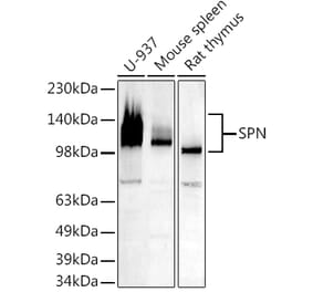
![Western Blot - Anti-CD43 Antibody [MEM-59] (A85600) - Antibodies.com](https://cdn.antibodies.com/image/catalog/85/A85600_94.jpg?profile=product_top)
![Immunohistochemistry - Anti-CD43 Antibody [MEM-59] (A85599) - Antibodies.com](https://cdn.antibodies.com/image/catalog/85/A85600_93.jpg?profile=product_top_thumb)
![Western Blot - Anti-CD43 Antibody [MEM-59] (A85600) - Antibodies.com](https://cdn.antibodies.com/image/catalog/85/A85600_94.jpg?profile=product_top_thumb)
![Immunohistochemistry - Anti-CD43 Antibody [MEM-59] (A85599) - Antibodies.com](https://cdn.antibodies.com/image/catalog/85/A85600_93.jpg?profile=product_image)
![Western Blot - Anti-CD43 Antibody [MEM-59] (A85600) - Antibodies.com](https://cdn.antibodies.com/image/catalog/85/A85600_94.jpg?profile=product_image)

![Immunohistochemistry - Anti-CD43 Antibody [rSPN/1094] - BSA and Azide free (A253211) - Antibodies.com](https://cdn.antibodies.com/image/catalog/253/A253211_1.jpg?profile=product_alternative)
![Immunohistochemistry - Anti-CD43 Antibody [DF-T1] - BSA and Azide free (A253206) - Antibodies.com](https://cdn.antibodies.com/image/catalog/253/A253206_1.jpg?profile=product_alternative)
![Immunohistochemistry - Anti-CD43 Antibody [SPN/839] (A250028) - Antibodies.com](https://cdn.antibodies.com/image/catalog/250/A250028_1.jpg?profile=product_alternative)
![Immunohistochemistry - Anti-CD43 Antibody [rSPN/1094] (A250031) - Antibodies.com](https://cdn.antibodies.com/image/catalog/250/A250031_1.jpg?profile=product_alternative)
![Immunohistochemistry - Anti-CD43 Antibody [SPN/839] - BSA and Azide free (A253208) - Antibodies.com](https://cdn.antibodies.com/image/catalog/253/A253208_1.jpg?profile=product_alternative)
![Immunohistochemistry - Anti-CD43 Antibody [DF-T1] (A250026) - Antibodies.com](https://cdn.antibodies.com/image/catalog/250/A250026_1.jpg?profile=product_alternative)
![Immunohistochemistry - Anti-CD43 Antibody [SPN/2049R] - BSA and Azide free (A253213) - Antibodies.com](https://cdn.antibodies.com/image/catalog/253/A253213_1.jpg?profile=product_alternative)
![Immunohistochemistry - Anti-CD43 Antibody [84-3C1] (A250029) - Antibodies.com](https://cdn.antibodies.com/image/catalog/250/A250029_1.jpg?profile=product_alternative)
![Immunohistochemistry - Anti-CD43 Antibody [SPN/3388] - BSA and Azide free (A253205) - Antibodies.com](https://cdn.antibodies.com/image/catalog/253/A253205_1.jpg?profile=product_alternative)
![Immunohistochemistry - Anti-CD43 Antibody [SPN/1766R] (A250032) - Antibodies.com](https://cdn.antibodies.com/image/catalog/250/A250032_1.jpg?profile=product_alternative)
![Immunohistochemistry - Anti-CD43 Antibody [SPN/1766R] - BSA and Azide free (A253212) - Antibodies.com](https://cdn.antibodies.com/image/catalog/253/A253212_1.jpg?profile=product_alternative)