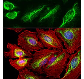
Immunofluorescent analysis of cortical neuron-glial cell culture from E20 rat stained with Anti-Vimentin Antibody, at a dilution of 1:5,000, in green, and Anti-GFAP Antibody (A85419 | 1:5,000, in red. The blue is DAPI staining of nuclear DNA. Fibroblastic cells contain only vimentin, so they are green. Developing astrocytic cells contain variable amounts of GFAP and vimentin, and so may appear red, green or yellow.
Western Blot - Anti-Vimentin Antibody [2A52] (A85423)
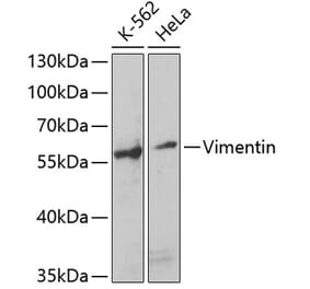
Western blot analysis of cell and whole brain tissue lysates using Anti-Vimentin Antibody, at a dilution of 1:5,000, in green,: [Lane 1] protein standard (red), [Lane 2] HEK293, [Lane 3] HeLa, [Lane 4] COS-1, [Lane 5] C6 cells, and [Lane 6] rat brain. The band at about 50 kDa mark corresponds to vimentin.
Western Blot - Anti-Vimentin Antibody [2A52] (A85423)
Western blot of crude extract of the human carcinoma HeLa cell line. Lane 18 was probed with Anti-Vimentin Antibody - showing a single sharp band at the expected molecular weight of 50 kDa. Lane 18 was probed with Anti-Vimentin Antibody (A85424). Lane 15 was probed with Anti-Actin Antibody (A85388) - giving an SDS-PAGE molecular weight of 42 kDa. Lane 16 was probed with Anti-14-3-3 eta Antibody (A85361) - which has an SDS-PAGE molecular weight of 28 kDa.
Immunohistochemistry analysis of a 4% PFA fixed paraffin embedded rat kidney section with Anti-Vimentin Antibody [2A52] (A85423) dilution 1:2,000 detected in DAB (brown) using the Vector Labs ImmPRESS method and reagents with citra buffer retrieval. Counterstained with Hematoxylin (blue). The Anti-Vimentin Antibody [2A52] (A85423) labels tubular epithelium in kidney. Note: this antibody performs well in testing with both 4% PFA and standard NBF fixed tissues but does not stain long term NBF fixed tissue effectively.
Publishing research using Anti-Vimentin Antibody [2A52] (A85423)? Please let us know so that we can list the citation on this page.


![Immunofluorescence - Anti-Vimentin Antibody [2A52] (A85423) - Antibodies.com](https://cdn.antibodies.com/image/catalog/85/A85423_1.jpg?profile=product_top)
![Western Blot - Anti-Vimentin Antibody [2A52] (A85423) - Antibodies.com](https://cdn.antibodies.com/image/catalog/85/A85423_2.jpg?profile=product_top)
![Western Blot - Anti-Vimentin Antibody [2A52] (A85423) - Antibodies.com](https://cdn.antibodies.com/image/catalog/85/A85423_3.jpg?profile=product_top)
![Immunohistochemistry - Anti-Vimentin Antibody [2A52] (A85423) - Antibodies.com](https://cdn.antibodies.com/image/catalog/85/A85423_4.jpg?profile=product_top)
![Immunofluorescence - Anti-Vimentin Antibody [2A52] (A85423) - Antibodies.com](https://cdn.antibodies.com/image/catalog/85/A85423_1.jpg?profile=product_top_thumb)
![Western Blot - Anti-Vimentin Antibody [2A52] (A85423) - Antibodies.com](https://cdn.antibodies.com/image/catalog/85/A85423_2.jpg?profile=product_top_thumb)
![Western Blot - Anti-Vimentin Antibody [2A52] (A85423) - Antibodies.com](https://cdn.antibodies.com/image/catalog/85/A85423_3.jpg?profile=product_top_thumb)
![Immunohistochemistry - Anti-Vimentin Antibody [2A52] (A85423) - Antibodies.com](https://cdn.antibodies.com/image/catalog/85/A85423_4.jpg?profile=product_top_thumb)
![Immunofluorescence - Anti-Vimentin Antibody [2A52] (A85423) - Antibodies.com](https://cdn.antibodies.com/image/catalog/85/A85423_1.jpg?profile=product_image)
![Western Blot - Anti-Vimentin Antibody [2A52] (A85423) - Antibodies.com](https://cdn.antibodies.com/image/catalog/85/A85423_2.jpg?profile=product_image)
![Western Blot - Anti-Vimentin Antibody [2A52] (A85423) - Antibodies.com](https://cdn.antibodies.com/image/catalog/85/A85423_3.jpg?profile=product_image)
![Immunohistochemistry - Anti-Vimentin Antibody [2A52] (A85423) - Antibodies.com](https://cdn.antibodies.com/image/catalog/85/A85423_4.jpg?profile=product_image)
![Immunocytochemistry - Anti-Vimentin Antibody [VI-10] (A86650) - Antibodies.com](https://cdn.antibodies.com/image/catalog/86/A86652_789.jpg?profile=product_alternative)
![Immunohistochemistry - Anti-Vimentin Antibody [VIM/1937R] - BSA and Azide free (A253494) - Antibodies.com](https://cdn.antibodies.com/image/catalog/253/A253494_1.jpg?profile=product_alternative)
![Western Blot - Anti-Vimentin Antibody [ARC0086] (A305554) - Antibodies.com](https://cdn.antibodies.com/image/catalog/305/A305554_1.jpg?profile=product_alternative)
![Immunohistochemistry - Anti-Vimentin Antibody [VIM/1937R] (A250314) - Antibodies.com](https://cdn.antibodies.com/image/catalog/250/A250314_1.jpg?profile=product_alternative)
![Immunohistochemistry - Anti-Vimentin Antibody [V9] (A250310) - Antibodies.com](https://cdn.antibodies.com/image/catalog/250/A250310_1.jpg?profile=product_alternative)
![Western Blot - Anti-Vimentin Antibody [VM452] (A250307) - Antibodies.com](https://cdn.antibodies.com/image/catalog/250/A250307_1.jpg?profile=product_alternative)
![Immunohistochemistry - Anti-Vimentin Antibody [V9] - BSA and Azide free (A253490) - Antibodies.com](https://cdn.antibodies.com/image/catalog/253/A253490_1.jpg?profile=product_alternative)

![Western Blot - Anti-Vimentin Antibody [VM452] - BSA and Azide free (A253487) - Antibodies.com](https://cdn.antibodies.com/image/catalog/253/A253487_1.jpg?profile=product_alternative)

![Immunohistochemistry - Anti-Vimentin Antibody [VIM/6576R] (A250313) - Antibodies.com](https://cdn.antibodies.com/image/catalog/250/A250313_1.jpg?profile=product_alternative)
![Immunohistochemistry - Anti-Vimentin Antibody [rVIM/6575] - BSA and Azide free (A253486) - Antibodies.com](https://cdn.antibodies.com/image/catalog/253/A253486_1.jpg?profile=product_alternative)