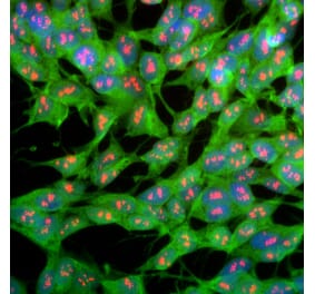
Immunofluorescent analysis of rat hippocampal section stained with Anti-UCH-L1 Antibody [BH7] (A85351), dilution 1:5,000, in green and co-stained with Anti-Fox3 Antibody (A85403), dilution 1:2,000, in red. The blue is Hoechst staining of nuclear DNA. Following transcardial perfusion of rat with 4% paraformaldehyde, the brain was post-fixed for 24 hours, cut to 45 µM, and free-floating sections were stained with the above antibodies. The UCH-L1 antibody stains the cell body and dendrites of hippocampal neurons, while the FOX3 antibody labels nuclei of the neuronal cells.
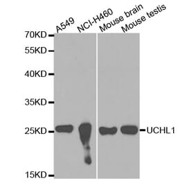
Western blot analysis of tissue lysates using Anti-UCH-L1 Antibody [BH7] (A85351), dilution 1:10,000, in green: [Lane 1] protein standard (red), [Lane 2] rat brain, [Lane 3] rat spinal cord, [Lane 4] mouse brain, [Lane 5] mouse spinal cord, [Lane 6] pig brain, and [Lane 7] pig spinal cord. The single band at 24 kDa corresponds to the UCH-L1 protein.
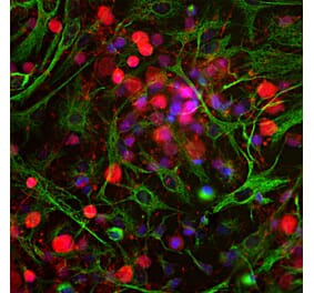
Immunofluorescent analysis of cortical neuron-glial culture from E20 rat stained with Anti-UCHL1 Antibody [BH7] (A85351) at a dilution of 1:5,000 (green) and costained with Anti-GFAP Antibody (A85307) at a dilution of 1:5,000 (red). Nuclei were stained with DAPI (blue). The Anti-UCHL1 Antibody [BH7] (A85351) antibody stains cell bodies and dendrites of neurons, while the GFAP antibody labels astrocytes.
A section of rat spinal cord stained with Anti-UCHL1 Antibody (red) and Anti-NF-H Antibody (A85336 | green). The large cells are a-motorneurons and UCHL1 fills the cytoplasm of their perikarya and dendrites. The Anti-NF-H Antibody binds primarily to phosphorylated axonal forms of NF-H, and so stains axons coursing between the large motor neurons.
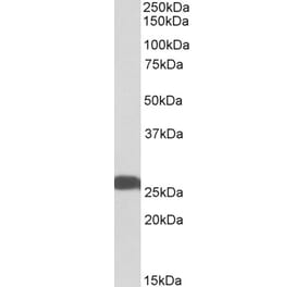
Blots of whole cell homogenate of rat and mouse brain stained with Anti-UCHL1 Antibody (Lanes 1 and 2 respectively). Lane S is a protein standard containing proteins of the indicated molecular weight. The antibody shows a major and clean band at about 26kDa. Image performed using an Odyssey infrared imager from Li-Cor Biosciences.
Immunohistochemistry analysis of a 4% PFA fixed paraffin embedded human brain cortex section with Anti-UCHL1 Antibody [BH7] (A85351) at a dilution of 1:10,000 detected in DAB (brown) following the ImmPress method with citra buffer retrieval. Counterstained with Hematoxylin (blue). UCHL1 is extremely abundant in the cell bodies and dendrites in a majority of neuronal cells. Note: this antibody performs well in testing with both 4% PFA and standard NBF fixed human, mouse and rat tissues.


![Immunofluorescence - Anti-UCHL1 Antibody [BH7] (A85351) - Antibodies.com](https://cdn.antibodies.com/image/catalog/85/A85351_1.jpg?profile=product_top)
![Western Blot - Anti-UCHL1 Antibody [BH7] (A85351) - Antibodies.com](https://cdn.antibodies.com/image/catalog/85/A85351_2.jpg?profile=product_top)
![Immunofluorescence - Anti-UCHL1 Antibody [BH7] (A85351) - Antibodies.com](https://cdn.antibodies.com/image/catalog/85/A85351_3.jpg?profile=product_top)
![Immunofluorescence - Anti-UCHL1 Antibody [BH7] (A85351) - Antibodies.com](https://cdn.antibodies.com/image/catalog/85/A85351_4.jpg?profile=product_top)
![Western Blot - Anti-UCHL1 Antibody [BH7] (A85351) - Antibodies.com](https://cdn.antibodies.com/image/catalog/85/A85351_5.jpg?profile=product_top)
![Immunohistochemistry - Anti-UCHL1 Antibody [BH7] (A85351) - Antibodies.com](https://cdn.antibodies.com/image/catalog/85/A85351_6.jpg?profile=product_top)
![Immunofluorescence - Anti-UCHL1 Antibody [BH7] (A85351) - Antibodies.com](https://cdn.antibodies.com/image/catalog/85/A85351_1.jpg?profile=product_top_thumb)
![Western Blot - Anti-UCHL1 Antibody [BH7] (A85351) - Antibodies.com](https://cdn.antibodies.com/image/catalog/85/A85351_2.jpg?profile=product_top_thumb)
![Immunofluorescence - Anti-UCHL1 Antibody [BH7] (A85351) - Antibodies.com](https://cdn.antibodies.com/image/catalog/85/A85351_3.jpg?profile=product_top_thumb)
![Immunofluorescence - Anti-UCHL1 Antibody [BH7] (A85351) - Antibodies.com](https://cdn.antibodies.com/image/catalog/85/A85351_4.jpg?profile=product_top_thumb)
![Western Blot - Anti-UCHL1 Antibody [BH7] (A85351) - Antibodies.com](https://cdn.antibodies.com/image/catalog/85/A85351_5.jpg?profile=product_top_thumb)
![Immunohistochemistry - Anti-UCHL1 Antibody [BH7] (A85351) - Antibodies.com](https://cdn.antibodies.com/image/catalog/85/A85351_6.jpg?profile=product_top_thumb)
![Immunofluorescence - Anti-UCHL1 Antibody [BH7] (A85351) - Antibodies.com](https://cdn.antibodies.com/image/catalog/85/A85351_1.jpg?profile=product_image)
![Western Blot - Anti-UCHL1 Antibody [BH7] (A85351) - Antibodies.com](https://cdn.antibodies.com/image/catalog/85/A85351_2.jpg?profile=product_image)
![Immunofluorescence - Anti-UCHL1 Antibody [BH7] (A85351) - Antibodies.com](https://cdn.antibodies.com/image/catalog/85/A85351_3.jpg?profile=product_image)
![Immunofluorescence - Anti-UCHL1 Antibody [BH7] (A85351) - Antibodies.com](https://cdn.antibodies.com/image/catalog/85/A85351_4.jpg?profile=product_image)
![Western Blot - Anti-UCHL1 Antibody [BH7] (A85351) - Antibodies.com](https://cdn.antibodies.com/image/catalog/85/A85351_5.jpg?profile=product_image)
![Immunohistochemistry - Anti-UCHL1 Antibody [BH7] (A85351) - Antibodies.com](https://cdn.antibodies.com/image/catalog/85/A85351_6.jpg?profile=product_image)
![Western Blot - Anti-PGP9.5 Antibody [13C4] (A250271) - Antibodies.com](https://cdn.antibodies.com/image/catalog/250/A250271_1.jpg?profile=product_alternative)
![Western Blot - Anti-PGP9.5 Antibody [ARC50367] (A307670) - Antibodies.com](https://cdn.antibodies.com/image/catalog/307/A307670_1.jpg?profile=product_alternative)
![Western Blot - Anti-PGP9.5 Antibody [13C4] - BSA and Azide free (A253451) - Antibodies.com](https://cdn.antibodies.com/image/catalog/253/A253451_1.jpg?profile=product_alternative)
![Immunohistochemistry - Anti-PGP9.5 Antibody [31A3] - BSA and Azide free (A253449) - Antibodies.com](https://cdn.antibodies.com/image/catalog/253/A253449_1.jpg?profile=product_alternative)
![Immunohistochemistry - Anti-PGP9.5 Antibody [31A3] (A250269) - Antibodies.com](https://cdn.antibodies.com/image/catalog/250/A250269_1.jpg?profile=product_alternative)
![Western Blot - Anti-PGP9.5 Antibody [SPM575] - BSA and Azide free (A253452) - Antibodies.com](https://cdn.antibodies.com/image/catalog/253/A253452_1.jpg?profile=product_alternative)
![Western Blot - Anti-PGP9.5 Antibody [SPM575] (A250272) - Antibodies.com](https://cdn.antibodies.com/image/catalog/250/A250272_1.jpg?profile=product_alternative)
![Immunohistochemistry - Anti-PGP9.5 Antibody [rUCHL1/775] - BSA and Azide free (A253455) - Antibodies.com](https://cdn.antibodies.com/image/catalog/253/A253455_1.jpg?profile=product_alternative)
![Immunohistochemistry - Anti-PGP9.5 Antibody [rUCHL1/775] (A250275) - Antibodies.com](https://cdn.antibodies.com/image/catalog/250/A250275_1.jpg?profile=product_alternative)
![Immunohistochemistry - Anti-PGP9.5 Antibody [SPM574] - BSA and Azide free (A253450) - Antibodies.com](https://cdn.antibodies.com/image/catalog/253/A253450_1.jpg?profile=product_alternative)