+1 (314) 370-6046 or
Contact Us - Argentina
- Australia
- Austria
- Bahrain
- Belgium
- Brazil
- Bulgaria
- Cameroon
- Canada
- Chile
- China
- Colombia
- Croatia
- Cyprus
- Czech Republic
- Denmark
- Ecuador
- Egypt
- Estonia
- Finland
- France
- Germany
- Greece
- Hong Kong
- Hungary
- Iceland
- India
- Indonesia
- Iran
- Ireland
- Israel
- Italy
- Japan
- Kazakhstan
- Kuwait
- Latvia
- Lithuania
- Luxembourg
- Macedonia
- Malaysia
- Malta
- Mexico
- Monaco
- Morocco
- Netherlands
- New Zealand
- Nigeria
- Norway
- Peru
- Philippines
- Poland
- Portugal
- Qatar
- Romania
- Russia
- Saudi Arabia
- Serbia
- Singapore
- Slovakia
- Slovenia
- South Africa
- South Korea
- Spain
- Sri Lanka
- Sweden
- Switzerland
- Taiwan
- Thailand
- Turkey
- Ukraine
- UAE
- United Kingdom
- United States
- Venezuela
- Vietnam


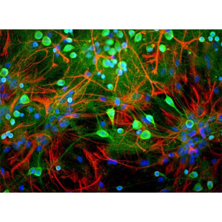
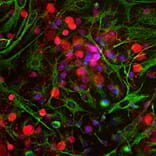
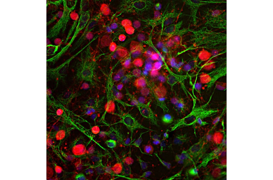
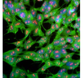
![Immunofluorescence - Anti-UCHL1 Antibody [BH7] (A85351) - Antibodies.com](https://cdn.antibodies.com/image/catalog/85/A85351_1.jpg?profile=product_alternative)
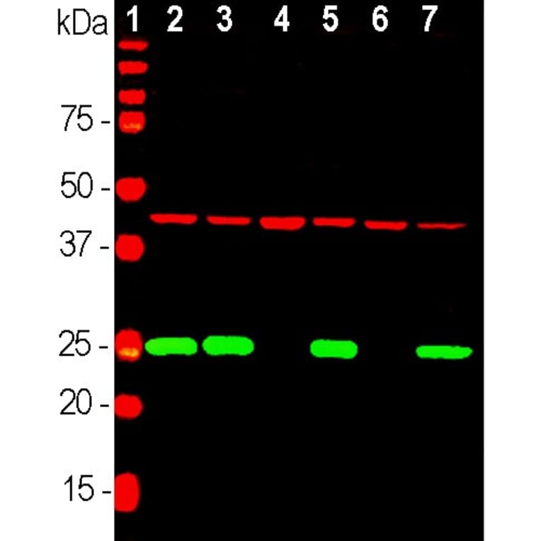
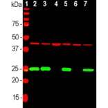
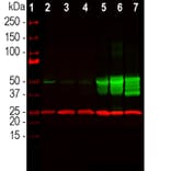
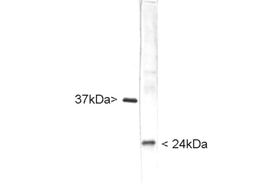
![Western Blot - Anti-PGP9.5 Antibody [SPM575] (A250272) - Antibodies.com](https://cdn.antibodies.com/image/catalog/250/A250272_1.jpg?profile=product_alternative)
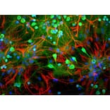
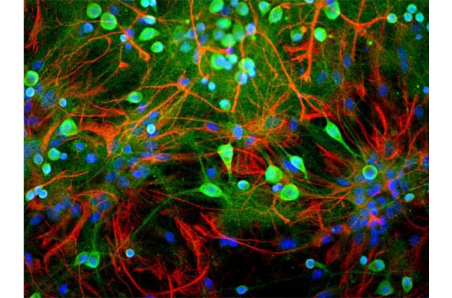
![Immunohistochemistry - Anti-PGP9.5 Antibody [SPM574] - BSA and Azide free (A253450) - Antibodies.com](https://cdn.antibodies.com/image/catalog/253/A253450_1.jpg?profile=product_alternative)
![Immunohistochemistry - Anti-PGP9.5 Antibody [SPM574] (A250270) - Antibodies.com](https://cdn.antibodies.com/image/catalog/250/A250270_1.jpg?profile=product_alternative)
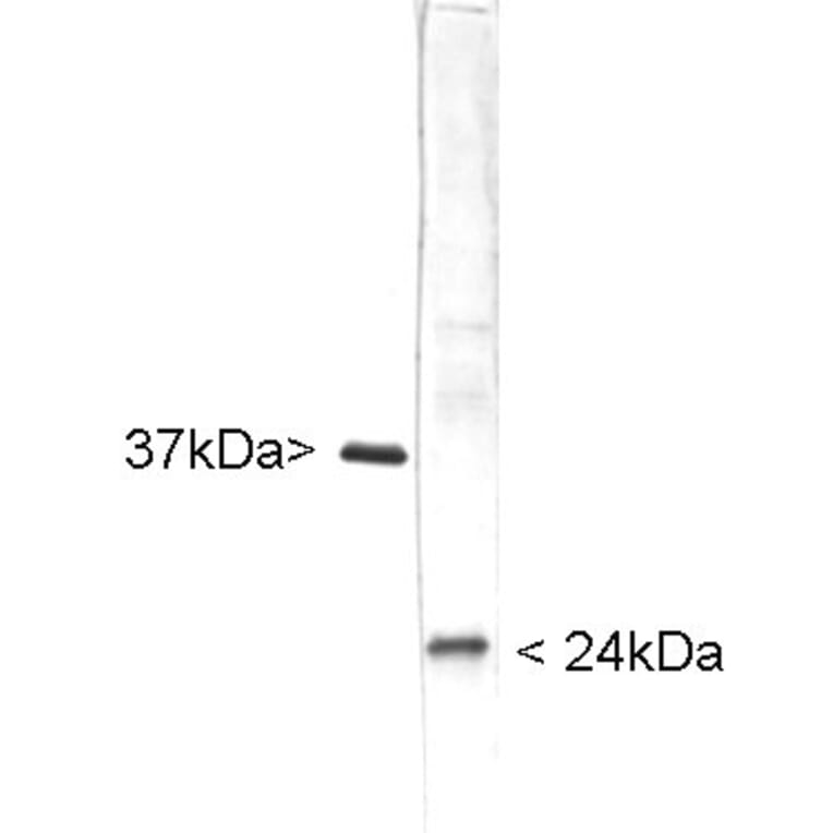
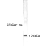
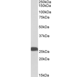
![Western Blot - Anti-PGP9.5 Antibody [13C4] (A250271) - Antibodies.com](https://cdn.antibodies.com/image/catalog/250/A250271_1.jpg?profile=product_alternative)
![Western Blot - Anti-PGP9.5 Antibody [13C4] - BSA and Azide free (A253451) - Antibodies.com](https://cdn.antibodies.com/image/catalog/253/A253451_1.jpg?profile=product_alternative)
![Immunohistochemistry - Anti-PGP9.5 Antibody [31A3] - BSA and Azide free (A253449) - Antibodies.com](https://cdn.antibodies.com/image/catalog/253/A253449_1.jpg?profile=product_alternative)
![Immunohistochemistry - Anti-PGP9.5 Antibody [31A3] (A250269) - Antibodies.com](https://cdn.antibodies.com/image/catalog/250/A250269_1.jpg?profile=product_alternative)
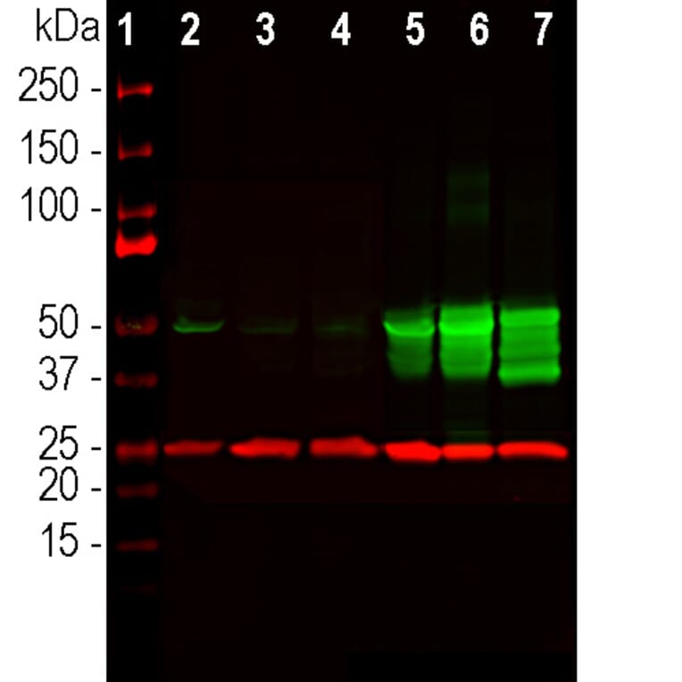
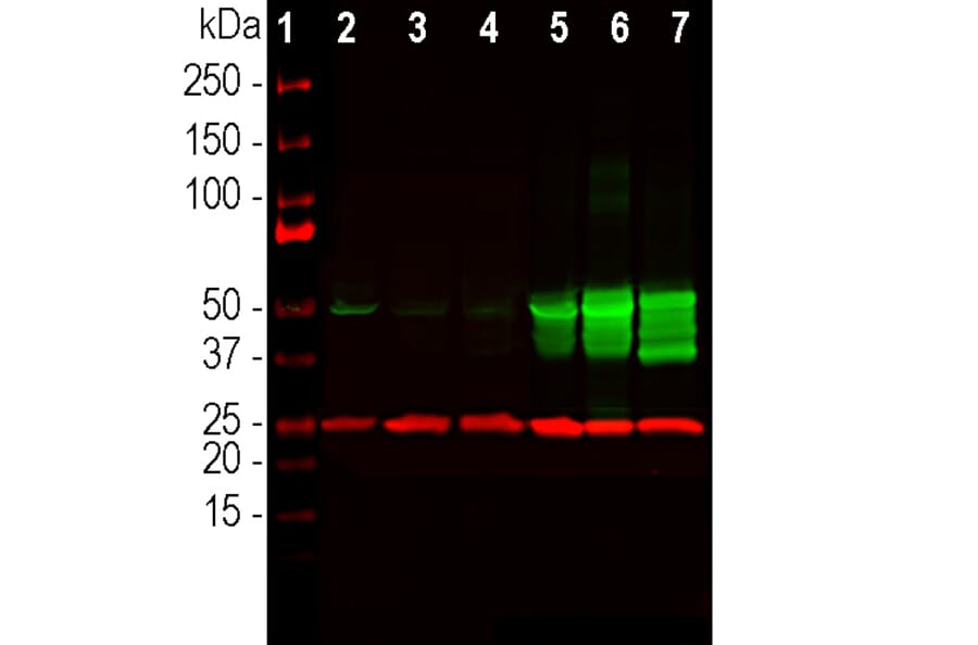
![Western Blot - Anti-PGP9.5 Antibody [ARC50367] (A307670) - Antibodies.com](https://cdn.antibodies.com/image/catalog/307/A307670_1.jpg?profile=product_alternative)
![Immunohistochemistry - Anti-PGP9.5 Antibody [rUCHL1/775] - BSA and Azide free (A253455) - Antibodies.com](https://cdn.antibodies.com/image/catalog/253/A253455_1.jpg?profile=product_alternative)
![Immunohistochemistry - Anti-PGP9.5 Antibody [rUCHL1/775] (A250275) - Antibodies.com](https://cdn.antibodies.com/image/catalog/250/A250275_1.jpg?profile=product_alternative)
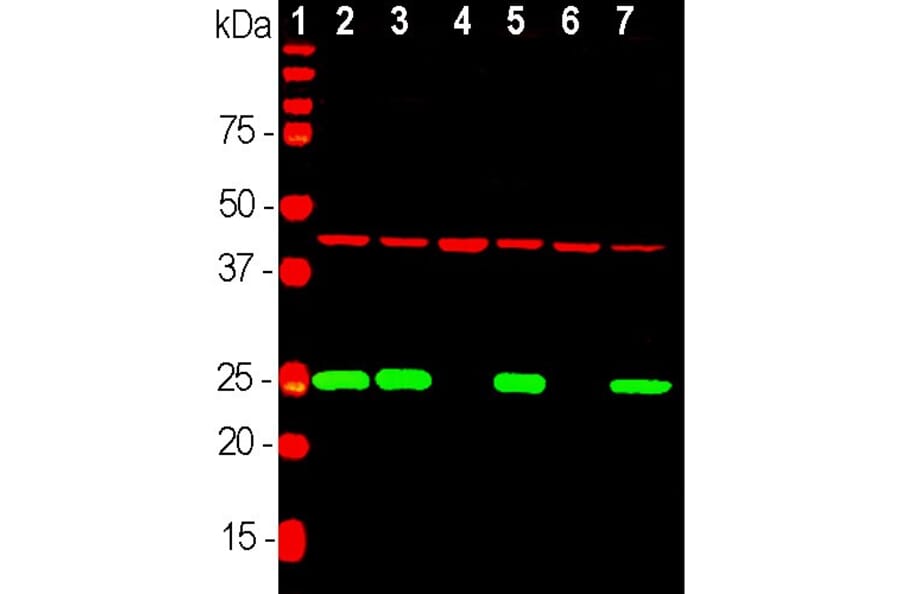
![Western Blot - Anti-PGP9.5 Antibody [SPM575] - BSA and Azide free (A253452) - Antibodies.com](https://cdn.antibodies.com/image/catalog/253/A253452_1.jpg?profile=product_alternative)