The epitope for this antibody is located in the peptide IQDKEGIPPDQQRLIFAGKQ, amino acids 30-49 of bovine Ubiquitin, with the central 10 amino acid segment, GIPPDQQRLI, likely to be the most significant component of this antibodies epitope.
Immunohistochemistry analysis of a NBF fixed paraffin embedded human hippocampus section from an Alzheimer’s Disease case. Mouse mAb to ubiquitin, Anti-Ubiquitin Antibody [Ubi-1] (A85456) at a dilution of 1:2,000, was detected in DAB (brown) following the ImmPress method with citra buffer retrieval. Counterstained with Hematoxylin (blue). The Anti-Ubiquitin Antibody [Ubi-1] (A85456) strongly labels flame shapped tangles in pyramidal neurons and dystrophic neurites nuclei characteristic of Alzheimer’s disease. Note: this antibody performs well in testing with 4% PFA or standard NBF fixed human and rat tissues.
Anti-Ubiquitin Antibody staining of cerebral cortex of an Alzheimer patient. Neurofibrillary tangles and dystrophic neurites associated with senile plaques stain strongly with this antibody. In the center is a typical neurofibrillary tangle containing neuron.
Immunohistochemistry analysis of a NBF fixed paraffin embedded human hippocampus section from an Alzheimer’s Disease case. Anti-Ubiquitin Antibody [Ubi-1] (A85456) at a dilution of 1:2,000 detected with DAB (brown) using the Vector Labs ImmPRESS method and reagents with citra buffer retrieval. Counterstained with Hematoxylin (blue). The Anti-Ubiquitin Antibody (A85455) antibody strongly labels the cytoplasm of diseased neurons as identified by their morphology.
Immunohistochemistry analysis of formalin fixed paraffin embedded cerebral cortex of an Alzheimer patient processed with Anti-Ubiquitin Antibody [Ubi-1] (A85456) using HRP/DAB (brown). Counterstained with Hematoxylin (blue). A typical flame shaped tangle is seen in a pyramidal neuron in the center and is surrounded by some dystrophic neurites, also strongly ubiquitin positive. Both are commonly seen in cortical and hippocampal Alzheimer brain sections and are typical for this disease, but are rare or absent in healthy brain.
Western Blot - Anti-Ubiquitin Antibody [Ubi-1] (A85456)
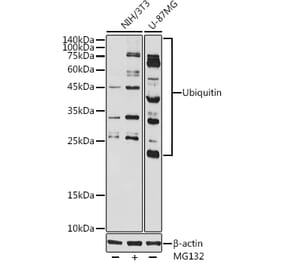
Blots of mono and polyubiquitin (Lane 1), monoubiquitin only (Lane 2), and 100 µg total wet weight of cerebellum, cortex and brain stem respectively (Lane 3-5) were probed with Anti-Ubiquitin Antibody. Material was run out on 20% SDS-PAGE and transferred electrophoretically to PVDF.
Western Blot - Anti-Ubiquitin Antibody [Ubi-1] (A85456)
Western blot analysis using Anti-Ubiquitin Antibody [Ubi-1] (A85456) of [Lane 1] mono and K48 linked polyubiquitin, [Lane 2] monoubiquitin only, [Lane 3] 100µg total wet weight of homogenates of rat cerebellum, [Lane 4] 100µg total wet weight of homogenates of rat cortex, and [Lane 5] 100µg total wet weight of homogenates of rat brain stem. Material was run out on 20% SDS-PAGE and transferred electrophoretically to PVDF. Anti-Ubiquitin Antibody [Ubi-1] (A85456) binds both mono and polyubiquitin and detects monoubiquitin in cell and tissue lysates.
Western Blot - Anti-Ubiquitin Antibody [Ubi-1] (A85456)
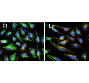
Western blot analysis of HEK293 cell lysates using Anti-Ubiquitin Antibody [Ubi-1] (A85456) at a dilution of 1:1,000 (green). The lanes contain: [Lane 1] protein standard (red), [Lane 2] cells maintained in normal medium, [Lane 3] cells treated with 10µM of proteasome inhibitor lactacystin (Lc) for 16hrs. Lysed cells were electrophoresed on 4-20% SDS-PAGE, and transferred to PVDF membranes. The smear detected above the 200kDa standard represent accumulation of ubiquitinated proteins in proteasome inhibitor-Lc treated cells. The prominent band at 8kDa corresponds to monoubiquitin. The same blot was probed with rabbit pAb to HSP60, RPCA-HSP60 at a dilution of 1:5,000 in red, used as a loading control.


![Immunohistochemistry - Anti-Ubiquitin Antibody [Ubi-1] (A85456) - Antibodies.com](https://cdn.antibodies.com/image/catalog/85/A85456_1.jpg?profile=product_top)
![Immunohistochemistry - Anti-Ubiquitin Antibody [Ubi-1] (A85456) - Antibodies.com](https://cdn.antibodies.com/image/catalog/85/A85456_2.jpg?profile=product_top)
![Immunohistochemistry - Anti-Ubiquitin Antibody [Ubi-1] (A85456) - Antibodies.com](https://cdn.antibodies.com/image/catalog/85/A85456_3.D.Hippocampus_Citra_20X.jpg?profile=product_top)
![Immunohistochemistry - Anti-Ubiquitin Antibody [Ubi-1] (A85456) - Antibodies.com](https://cdn.antibodies.com/image/catalog/85/A85456_4.jpg?profile=product_top)
![Western Blot - Anti-Ubiquitin Antibody [Ubi-1] (A85456) - Antibodies.com](https://cdn.antibodies.com/image/catalog/85/A85456_5.jpg?profile=product_top)
![Western Blot - Anti-Ubiquitin Antibody [Ubi-1] (A85456) - Antibodies.com](https://cdn.antibodies.com/image/catalog/85/A85456_6.jpg?profile=product_top)
![Western Blot - Anti-Ubiquitin Antibody [Ubi-1] (A85456) - Antibodies.com](https://cdn.antibodies.com/image/catalog/85/A85456_7.jpg?profile=product_top)
![Immunohistochemistry - Anti-Ubiquitin Antibody [Ubi-1] (A85456) - Antibodies.com](https://cdn.antibodies.com/image/catalog/85/A85456_1.jpg?profile=product_top_thumb)
![Immunohistochemistry - Anti-Ubiquitin Antibody [Ubi-1] (A85456) - Antibodies.com](https://cdn.antibodies.com/image/catalog/85/A85456_2.jpg?profile=product_top_thumb)
![Immunohistochemistry - Anti-Ubiquitin Antibody [Ubi-1] (A85456) - Antibodies.com](https://cdn.antibodies.com/image/catalog/85/A85456_3.D.Hippocampus_Citra_20X.jpg?profile=product_top_thumb)
![Immunohistochemistry - Anti-Ubiquitin Antibody [Ubi-1] (A85456) - Antibodies.com](https://cdn.antibodies.com/image/catalog/85/A85456_4.jpg?profile=product_top_thumb)
![Western Blot - Anti-Ubiquitin Antibody [Ubi-1] (A85456) - Antibodies.com](https://cdn.antibodies.com/image/catalog/85/A85456_5.jpg?profile=product_top_thumb)
![Western Blot - Anti-Ubiquitin Antibody [Ubi-1] (A85456) - Antibodies.com](https://cdn.antibodies.com/image/catalog/85/A85456_6.jpg?profile=product_top_thumb)
![Western Blot - Anti-Ubiquitin Antibody [Ubi-1] (A85456) - Antibodies.com](https://cdn.antibodies.com/image/catalog/85/A85456_7.jpg?profile=product_top_thumb)
![Immunohistochemistry - Anti-Ubiquitin Antibody [Ubi-1] (A85456) - Antibodies.com](https://cdn.antibodies.com/image/catalog/85/A85456_1.jpg?profile=product_image)
![Immunohistochemistry - Anti-Ubiquitin Antibody [Ubi-1] (A85456) - Antibodies.com](https://cdn.antibodies.com/image/catalog/85/A85456_2.jpg?profile=product_image)
![Immunohistochemistry - Anti-Ubiquitin Antibody [Ubi-1] (A85456) - Antibodies.com](https://cdn.antibodies.com/image/catalog/85/A85456_3.D.Hippocampus_Citra_20X.jpg?profile=product_image)
![Immunohistochemistry - Anti-Ubiquitin Antibody [Ubi-1] (A85456) - Antibodies.com](https://cdn.antibodies.com/image/catalog/85/A85456_4.jpg?profile=product_image)
![Western Blot - Anti-Ubiquitin Antibody [Ubi-1] (A85456) - Antibodies.com](https://cdn.antibodies.com/image/catalog/85/A85456_5.jpg?profile=product_image)
![Western Blot - Anti-Ubiquitin Antibody [Ubi-1] (A85456) - Antibodies.com](https://cdn.antibodies.com/image/catalog/85/A85456_6.jpg?profile=product_image)
![Western Blot - Anti-Ubiquitin Antibody [Ubi-1] (A85456) - Antibodies.com](https://cdn.antibodies.com/image/catalog/85/A85456_7.jpg?profile=product_image)
![Immunocytochemistry/Immunofluorescence - Anti-Ubiquitin Antibody [FK2] (A305218) - Antibodies.com](https://cdn.antibodies.com/image/catalog/305/A305218_1.png?profile=product_alternative)
![Western Blot - Anti-Ubiquitin Antibody [ARC50024] (A307666) - Antibodies.com](https://cdn.antibodies.com/image/catalog/307/A307666_1.jpg?profile=product_alternative)
![Immunohistochemistry - Anti-Ubiquitin Antibody [UBB/1748] - BSA and Azide free (A253441) - Antibodies.com](https://cdn.antibodies.com/image/catalog/253/A253441_1.jpg?profile=product_alternative)
![Immunohistochemistry - Anti-Ubiquitin Antibody [UBB/1748] (A250261) - Antibodies.com](https://cdn.antibodies.com/image/catalog/250/A250261_1.jpg?profile=product_alternative)
![Western Blot - Anti-Ubiquitin Antibody [5B9-B3] (A305078) - Antibodies.com](https://cdn.antibodies.com/image/catalog/305/A305078_1.png?profile=product_alternative)
![Immunocytochemistry/Immunofluorescence - Anti-Ubiquitin Antibody [6C11-B3] (A305079) - Antibodies.com](https://cdn.antibodies.com/image/catalog/305/A305079_1.png?profile=product_alternative)
![Immunohistochemistry - Anti-Ubiquitin Antibody [UBB/2122] - BSA and Azide free (A253442) - Antibodies.com](https://cdn.antibodies.com/image/catalog/253/A253442_1.jpg?profile=product_alternative)