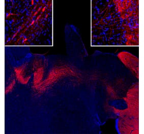
Immunofluorescent analysis of rat brain section stained with Anti-Tyrosine Hydroxylase Antibody [4H2] (A104315), at a dilution of 1:1,000, in red. The blue is Hoechst staining of nuclear DNA. Following transcardial perfusion of rat with 4% paraformaldehyde, brain was post fixed for 24 hours, cut to 45µM, and free-floating sections were stained. Anti-Tyrosine Hydroxylase Antibody [4H2] (A104315) stains Tyrosine Hydroxylase expressing neuronal processes, which are particularly numerous in the striatum, at the right of the image.
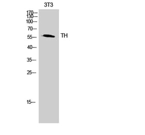
Western Blot - Anti-Tyrosine Hydroxylase Antibody [4H2] (A104315)
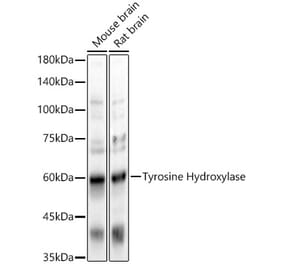
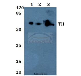
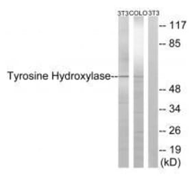
Western blot analysis of tissue and cell lysates using Anti-Tyrosine Hydroxylase Antibody [4H2] (A104315), at a dilution of 1:5,000, in green. The lanes contain: [Lane 1] protein standard (red), [Lane 2] rat brain caudate/putmen, and [Lane 3] PC12 cells. The strong band at about 58kDa corresponds to Tyrosine Hydroxylase protein. The weak band at about 80kDa in the rat brain homogenate is of unknown origin, but does not affect the specific cell and process staining of this antibody.
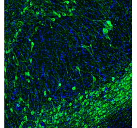
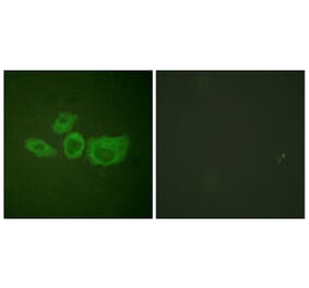
A section of mouse midbrain stained with Anti-Tyrosine Hydroxylase Antibody [4H2] (A104315) (green). The cytoplasm and processes of these dopaminergic neurons are revealed.
Immunohistochemistry analysis of a 4% PFA fixed paraffin embedded rat brain section with Anti-Tyrosine Hydroxylase Antibody [4H2] (A104315) at a dilution of 1:2,000. Anti-Tyrosine Hydroxylase Antibody [4H2] (A104315) strongly labels chatecholaminergic neurons within the substantia nigra. Note: Note: this antibody has not been tested in NBF fixed material.
Publishing research using Anti-Tyrosine Hydroxylase Antibody [4H2] (A104315)? Please let us know so that we can list the citation on this page.
Alternative products to Anti-Tyrosine Hydroxylase Antibody [4H2] (A104315)


![Immunofluorescence - Anti-Tyrosine Hydroxylase Antibody [4H2] (A104315) - Antibodies.com](https://cdn.antibodies.com/image/catalog/104/A104315_1.jpg?profile=product_top)
![Western Blot - Anti-Tyrosine Hydroxylase Antibody [4H2] (A104315) - Antibodies.com](https://cdn.antibodies.com/image/catalog/104/A104315_2.jpg?profile=product_top)
![Immunofluorescence - Anti-Tyrosine Hydroxylase Antibody [4H2] (A104315) - Antibodies.com](https://cdn.antibodies.com/image/catalog/104/A104315_3.jpg?profile=product_top)
![Immunohistochemistry - Anti-Tyrosine Hydroxylase Antibody [4H2] (A104315) - Antibodies.com](https://cdn.antibodies.com/image/catalog/104/A104315_4.jpg?profile=product_top)
![Immunofluorescence - Anti-Tyrosine Hydroxylase Antibody [4H2] (A104315) - Antibodies.com](https://cdn.antibodies.com/image/catalog/104/A104315_1.jpg?profile=product_top_thumb)
![Western Blot - Anti-Tyrosine Hydroxylase Antibody [4H2] (A104315) - Antibodies.com](https://cdn.antibodies.com/image/catalog/104/A104315_2.jpg?profile=product_top_thumb)
![Immunofluorescence - Anti-Tyrosine Hydroxylase Antibody [4H2] (A104315) - Antibodies.com](https://cdn.antibodies.com/image/catalog/104/A104315_3.jpg?profile=product_top_thumb)
![Immunohistochemistry - Anti-Tyrosine Hydroxylase Antibody [4H2] (A104315) - Antibodies.com](https://cdn.antibodies.com/image/catalog/104/A104315_4.jpg?profile=product_top_thumb)
![Immunofluorescence - Anti-Tyrosine Hydroxylase Antibody [4H2] (A104315) - Antibodies.com](https://cdn.antibodies.com/image/catalog/104/A104315_1.jpg?profile=product_image)
![Western Blot - Anti-Tyrosine Hydroxylase Antibody [4H2] (A104315) - Antibodies.com](https://cdn.antibodies.com/image/catalog/104/A104315_2.jpg?profile=product_image)
![Immunofluorescence - Anti-Tyrosine Hydroxylase Antibody [4H2] (A104315) - Antibodies.com](https://cdn.antibodies.com/image/catalog/104/A104315_3.jpg?profile=product_image)
![Immunohistochemistry - Anti-Tyrosine Hydroxylase Antibody [4H2] (A104315) - Antibodies.com](https://cdn.antibodies.com/image/catalog/104/A104315_4.jpg?profile=product_image)