Unconjugated
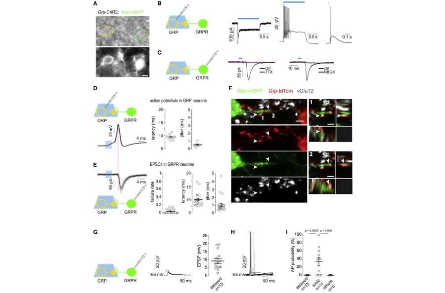
Spinal transmission of pruritoceptive (itch) signals requires transneuronal signaling by gastrin-releasing peptide (GRP) produced by a subpopulation of dorsal horn excitatory interneurons. These neurons also express the glutamatergic marker vGluT2, raising the question of why glutamate alone is insufficient for spinal itch relay. Using optogenetics together with slice electrophysiology and mouse behavior, we demonstrate that baseline synaptic coupling between GRP and GRP receptor (GRPR) neurons is too weak for suprathreshold excitation. Only when we mimicked the endogenous firing of GRP neurons and stimulated them repetitively to fire bursts of action potentials did GRPR neurons depolarize progressively and become excitable by GRP neurons. GRPR but not glutamate receptor antagonism prevented this action. Provoking itch-like behavior by optogenetic activation of spinal GRP neurons required similar stimulation paradigms. These results establish a spinal gating mechanism for itch that requires sustained repetitive activity of presynaptic GRP neurons and postsynaptic GRP signaling to drive GRPR neuron output.
Copyright © 2019 The Author(s). Published by Elsevier Inc. All rights reserved.
Oligodendrocytes produce myelin for rapid transmission and saltatory conduction of action potentials in the vertebrate central nervous system. Activation of the myelination program requires several transcription factors including Sox10, Olig2, and Nkx2.2. Functional interactions among them are poorly understood and important components of the regulatory network are still unknown. Here, we identify Nfat proteins as Sox10 targets and regulators of oligodendroglial differentiation in rodents and humans. Overall levels and nuclear fraction increase during differentiation. Inhibition of Nfat activity impedes oligodendrocyte differentiation in vitro and in vivo. On a molecular level, Nfat proteins cooperate with Sox10 to relieve reciprocal repression of Olig2 and Nkx2.2 as precondition for oligodendroglial differentiation and myelination. As Nfat activity depends on calcium-dependent activation of calcineurin signaling, regulatory network and oligodendroglial differentiation become sensitive to calcium signals. NFAT proteins are also detected in human oligodendrocytes, downregulated in active multiple sclerosis lesions and thus likely relevant in demyelinating disease.
ATOH1 is a master transcription factor for the secretory lineage differentiation of intestinal epithelial cells (IECs). However, the comprehensive contribution of ATOH1+ secretory lineage IECs to the homeostasis, repair, and tumorigenesis of the intestinal epithelium remains uncertain. Through our ATOH1+ cell-lineage tracing, we show here that a definite number of ATOH1+ IECs retain stem cell properties and can form ATOH1+IEC-derived clonal ribbons (ATOH1+ICRs) under completely homeostatic conditions. Interestingly, colonic ATOH1+ IECs appeared to exhibit their stem cell function more frequently compared with those of the small intestine. Consistently, the formation of ATOH1+ICRs was significantly enhanced upon dextran sodium sulfate colitis-induced mucosal damage. In addition, colonic ATOH1+ IECs acquired tumor stem cell-like properties in the azoxymethane-DSS tumor model. Our results reveal an unexpected contribution of colonic ATOH1+ IECs to maintaining the stem cell population under both homeostatic and pathologic conditions and further illustrate the high plasticity of the crypt-intrinsic stem cell hierarchy.
Copyright © 2017 The Author(s). Published by Elsevier Inc. All rights reserved.
Proper function of the neural network results from the precise connections between axons and dendrites of presynaptic and postsynaptic neurons, respectively. In the Drosophila olfactory system, the dendrites of projection neurons (PNs) stereotypically target one of ~50 glomeruli in the antennal lobe (AL), the primary olfactory center in the brain, and form synapses with the axons of olfactory receptor neurons (ORNs). Here, we show that Eph and Ephrin, the well-known axon guidance molecules, instruct the dendrodendritic segregation during the discrete olfactory map formation. The Eph receptor tyrosine kinase is highly expressed and localized in the glomeruli related to reproductive behavior in the developing AL. In one of the pheromone-sensing glomeruli (DA1), the Eph cell-autonomously regulates its dendrites to reside in a single glomerulus by interacting with Ephrins expressed in adjacent PN dendrites. Our data demonstrate that the trans interaction between dendritic Eph and Ephrin is essential for the PN dendritic boundary formation in the DA1 olfactory circuit, potentially enabling strict segregation of odor detection between pheromones and the other odors.
© 2017 Anzo et al.; Published by Cold Spring Harbor Laboratory Press.
Malaria parasites, transmitted by the bite of an anopheline mosquito, pose an immense public health burden on many tropical and subtropical regions. The most important malaria vectors in sub-Saharan Africa are mosquitoes of the Anopheles gambiae complex including An. gambiae (sensu stricto). Given the increasing rates of insecticide resistance in these mosquitoes, alternative control strategies based on the release of genetically modified males are being evaluated to stop transmission by these disease vectors. These strategies rely on the mating competitiveness of release males, however currently there is no method to determine male mating success without sacrificing the female. Interestingly, unlike other insects, during mating An. gambiae males transfer their male accessory glands (MAGs) seminal secretions as a coagulated mating plug which is deposited in the female atrium.
Here we exploit this male reproductive feature and validate the use of a MAG-specific promoter to fluorescently label the mating plug and visualize the occurrence of insemination in vivo. We used the promoter region of the major mating plug protein, Plugin, to control the expression of a Plugin-tdTomato (PluTo) fusion protein, hypothesizing that this fusion protein could be incorporated into the plug for sexual transfer to the female. Anopheles gambiae PluTo transgenic males showed strong red fluorescence specifically in the MAGs and with a pattern closely matching endogenous Plugin expression. Moreover, the fusion protein was integrated into the mating plug and transferred to the female atrium during mating where it could be visualized microscopically in vivo without sacrificing the female. PluTo males were equally as competitive at mating as wild type males, and females mated to these males did not show any reduction in reproductive fitness.
The validation of the first MAG-specific promoter in transgenic An. gambiae facilitates the live detection of successful insemination hours after copulation has occurred. This provides a valuable tool for the assessment of male mating competitiveness not only in laboratory experiments but also in semi-field and field studies aimed at testing the feasibility of releasing genetically modified mosquitoes for disease control.
Causal relationships between central cardiovascular pathways and sympathetic vasomotor tone have not been evidenced. This study aimed to verify the sympathoexcitatory role of hypothalamic paraventricular nucleus neurons that project to the rostral ventrolateral medulla (PVN-RVLM neurons). By using optogenetic techniques, we demonstrated that stimulation of PVN-RVLM glutamatergic neurons increased renal sympathetic nerve activity and arterial pressure via, at least in part, stimulation of RVLM C1 neurons in rats. This monosynaptic pathway may function in acute sympathetic adjustments to stressors and/or be a component of chronic sympathetic hyperactivity in pathological conditions such as heart failure.
The rostral ventrolateral medulla (RVLM), which is known to play an important role in regulating sympathetic vasomotor tone, receives axonal projections from the hypothalamic paraventricular nucleus (PVN). However, no studies have proved that excitation of the PVN neurons that send axonal projections to the RVLM (PVN-RVLM neurons) causes sympathoexcitation. This study aimed to directly examine the sympathoexcitatory role of PVN-RVLM neurons. Male rats received microinjections into the PVN with an adeno-associated virus (AAV) vector that encoded a hybrid of channelrhodopsin-2/1 with the reporter tdTomato (ChIEF-tdTomato), or into the RVLM with a retrograde AAV vector that encoded a channelrhodopsin with green fluorescent protein (ChR2-GFPretro ). Under anaesthesia with urethane and a-chloralose, photostimulation (473 nm wavelength) of PVN-RVLM neurons, achieved by laser illumination of either RVLM of ChIEF-tdTomato rats (n = 8) or PVN of ChR2-GFPretro rats (n = 4), elicited significant renal sympathoexcitation. Immunofluorescence confocal microscopy showed that RVLM adrenergic C1 neurons of ChIEF-tdTomato rats were closely associated with tdTomato-labelled, PVN-derived axons that contained vesicular glutamate transporter 2. In another subset of anaesthetized ChIEF-tdTomato rats (n = 6), the renal sympathoexcitation elicited by photostimulation of the PVN was suppressed by administering ionotropic glutamate receptor blockers into the RVLM. These results demonstrate that excitation of PVN-RVLM glutamatergic neurons leads to sympathoexcitation via, at least in part, stimulation of RVLM C1 neurons.
© 2018 The Authors. The Journal of Physiology © 2018 The Physiological Society.
Variations in the human FTO gene have been linked to obesity and altered connectivity of the dopaminergic neurocircuitry. Here, we report that fat mass and obesity-associated protein (FTO) in dopamine D2 receptor-expressing medium spiny neurons (D2 MSNs) of mice regulate the excitability of these cells and control their striatopallidal globus pallidus external (GPe) projections. Lack of FTO in D2 MSNs translates into increased locomotor activity to novelty, associated with altered timing behavior, without impairing the ability to control actions or affecting reward-driven and conditioned behavior. Pharmacological manipulations of dopamine D1 receptor (D1R)- or D2R-dependent pathways in these animals reveal altered responses to D1- and D2-MSN-mediated control of motor output. These findings reveal a critical role for FTO to control D2 MSN excitability, their projections to the GPe, and behavioral responses to novelty.
Copyright © 2019 The Authors. Published by Elsevier Inc. All rights reserved.
While it is now appreciated that certain long noncoding RNAs (lncRNAs) have important functions in cell biology, relatively few have been shown to regulate development in vivo, particularly with genetic strategies that establish cis versus trans mechanisms. Pnky is a nuclear-enriched lncRNA that is transcribed divergently from the neighboring proneural transcription factor Pou3f2. Here, we show that conditional deletion of Pnky from the developing cortex regulates the production of projection neurons from neural stem cells (NSCs) in a cell-autonomous manner, altering postnatal cortical lamination. Surprisingly, Pou3f2 expression is not disrupted by deletion of the entire Pnky gene. Moreover, expression of Pnky from a BAC transgene rescues the differential gene expression and increased neurogenesis of Pnky-knockout NSCs, as well as the developmental phenotypes of Pnky-deletion in vivo. Thus, despite being transcribed divergently from a key developmental transcription factor, the lncRNA Pnky regulates development in trans.
Published by Elsevier Inc.
Neuropeptide Y (NPY) exerts a powerful orexigenic effect in the hypothalamus. However, extra-hypothalamic nuclei also produce NPY, but its influence on energy homeostasis is unclear. Here we uncover a previously unknown feeding stimulatory pathway that is activated under conditions of stress in combination with calorie-dense food; NPY neurons in the central amygdala are responsible for an exacerbated response to a combined stress and high-fat-diet intervention. Central amygdala NPY neuron-specific Npy overexpression mimics the obese phenotype seen in a combined stress and high-fat-diet model, which is prevented by the selective ablation of Npy. Using food intake and energy expenditure as readouts, we demonstrate that selective activation of central amygdala NPY neurons results in increased food intake and decreased energy expenditure. Mechanistically, it is the diminished insulin signaling capacity on central amygdala NPY neurons under combined stress and high-fat-diet conditions that leads to the exaggerated development of obesity.
Copyright © 2019 Elsevier Inc. All rights reserved.
Among the diverse interneuron subtypes in the neocortex, chandelier cells (ChCs) are the only population that selectively innervate pyramidal neurons (PyNs) at their axon initial segment (AIS), the site of action potential initiation, allowing them to exert powerful control over PyN output. Yet, mechanisms underlying their subcellular innervation of PyN AISs are unknown. To identify molecular determinants of ChC/PyN AIS innervation, we performed an in vivo RNAi screen of PyN-expressed axonal cell adhesion molecules (CAMs) and select Ephs/ephrins. Strikingly, we found the L1 family member L1CAM to be the only molecule required for ChC/PyN AIS innervation. Further, we show that L1CAM is required during both the establishment and maintenance of innervation, and that selective innervation of PyN AISs by ChCs requires AIS anchoring of L1CAM by the cytoskeletal ankyrin-G/ßIV-spectrin complex. Thus, our findings identify PyN-expressed L1CAM as a critical CAM required for innervation of neocortical PyN AISs by ChCs. VIDEO ABSTRACT.
Copyright © 2019 Elsevier Inc. All rights reserved.
Gastrin-releasing peptide (GRP) is a spinal itch transmitter expressed by a small population of dorsal horn interneurons (GRP neurons). The contribution of these neurons to spinal itch relay is still only incompletely understood, and their potential contribution to pain-related behaviors remains controversial. Here, we have addressed this question in a series of experiments performed in GRP::cre and GRP::eGFP transgenic male mice. We combined behavioral tests with neuronal circuit tracing, morphology, chemogenetics, optogenetics, and electrophysiology to obtain a more comprehensive picture. We found that GRP neurons form a rather homogeneous population of central cell-like excitatory neurons located in lamina II of the superficial dorsal horn. Multicolor high-resolution confocal microscopy and optogenetic experiments demonstrated that GRP neurons receive direct input from MrgprA3-positive pruritoceptors. Anterograde HSV-based neuronal tracing initiated from GRP neurons revealed ascending polysynaptic projections to distinct areas and nuclei in the brainstem, midbrain, thalamus, and the somatosensory cortex. Spinally restricted ablation of GRP neurons reduced itch-related behaviors to different pruritogens, whereas their chemogenetic excitation elicited itch-like behaviors and facilitated responses to several pruritogens. By contrast, responses to painful stimuli remained unaltered. These data confirm a critical role of dorsal horn GRP neurons in spinal itch transmission but do not support a role in pain.SIGNIFICANCE STATEMENT Dorsal horn gastrin-releasing peptide neurons serve a well-established function in the spinal transmission of pruritic (itch) signals. A potential role in the transmission of nociceptive (pain) signals has remained controversial. Our results provide further support for a critical role of dorsal horn gastrin-releasing peptide neurons in itch circuits, but we failed to find evidence supporting a role in pain.
Copyright © 2019 the authors 0270-6474/19/392238-13$15.00/0.
The establishment of axon-dendrite polarity is fundamental for radial migration of neurons during cortex development of mammals. We demonstrate that the E3 ubiquitin ligases WW-Containing Proteins 1 and 2 (Wwp1 and Wwp2) are indispensable for proper polarization of developing neurons. We show that knockout of Wwp1 and Wwp2 results in defects in axon-dendrite polarity in pyramidal neurons, and their aberrant laminar cortical distribution. Knockout of miR-140, encoded in Wwp2 intron, engenders phenotypic changes analogous to those upon Wwp1 and Wwp2 deletion. Intriguingly, transcription of the Wwp1 and Wwp2/miR-140 loci in neurons is induced by the transcription factor Sox9. Finally, we provide evidence that miR-140 supervises the establishment of axon-dendrite polarity through repression of Fyn kinase mRNA. Our data delineate a novel regulatory pathway that involves Sox9-[Wwp1/Wwp2/miR-140]-Fyn required for axon specification, acquisition of pyramidal morphology, and proper laminar distribution of cortical neurons.
Copyright © 2018 Elsevier Inc. All rights reserved.
Understanding the migration of newborn neurons within the brain presents a major challenge in contemporary biology. Neuronal migration is widespread within the developing brain but is also important within the adult brain. For instance, stem cells within the ventricular-subventricular zone (V-SVZ) and the subgranular zone of dentate gyrus of the adult rodent brain produce neuroblasts that migrate to the olfactory bulb and granule cell layer of the dentate gyrus, respectively, where they regulate key brain functions including innate olfactory responses, learning, and memory. Critically, our understanding of the factors mediating neuroblast migration remains limited. The transcription factor nuclear factor I X (NFIX) has previously been implicated in embryonic cortical development. Here, we employed conditional ablation of Nfix from the adult mouse brain and demonstrated that the removal of this gene from either neural stem and progenitor cells, or neuroblasts, within the V-SVZ culminated in neuroblast migration defects. Mechanistically, we identified aberrant neuroblast branching, due in part to increased expression of the guanylyl cyclase natriuretic peptide receptor 2 (Npr2), as a factor contributing to abnormal migration in Nfix-deficient adult mice. Collectively, these data provide new insights into how neuroblast migration is regulated at a transcriptional level within the adult brain.
© The Author(s) 2018. Published by Oxford University Press. All rights reserved. For Permissions, please e-mail: journals.permissions@oup.com.
Acute infection, if not kept in check, can lead to systemic inflammatory responses in the brain. Here, we show that within 2 hr of systemic inflammation, PDGFRß mural cells of blood vessels rapidly secrete chemokine CCL2, which in turn increases total neuronal excitability by promoting excitatory synaptic transmission in glutamatergic neurons of multiple brain regions. By single-cell RNA sequencing, we identified Col1a1 and Rgs5 subgroups of PDGFRß cells as the main source of CCL2. Lipopolysaccharide (LPS)- or Poly(I:C)-treated pericyte culture medium induced similar effects in a CCL2-dependent manner. Importantly, in Pdgfrb-Cre;Ccl2fl/fl mice, LPS-induced increase in excitatory synaptic transmission was significantly attenuated. These results demonstrate in vivo that PDGFRß cells function as initial sensors of external insults by secreting CCL2, which relays the signal to the central nervous system. Through their gateway position in the brain, PDGFRß cells are ideally positioned to respond rapidly to environmental changes and to coordinate responses.
Copyright © 2018 Elsevier Inc. All rights reserved.
Delineating the basic cellular components of cortical inhibitory circuits remains a fundamental issue in order to understand their specific contributions to microcircuit function. It is still unclear how current classifications of cortical interneuron subtypes relate to biological processes such as their developmental specification. Here we identified the developmental trajectory of neurogliaform cells (NGCs), the main effectors of a powerful inhibitory motif recruited by long-range connections. Using in vivo genetic lineage-tracing in mice, we report that NGCs originate from a specific pool of 5-HT3AR-expressing Hmx3+ cells located in the preoptic area (POA). Hmx3-derived 5-HT3AR+ cortical interneurons (INs) expressed the transcription factors PROX1, NR2F2, the marker reelin but not VIP and exhibited the molecular, morphological and electrophysiological profile of NGCs. Overall, these results indicate that NGCs are a distinct class of INs with a unique developmental trajectory and open the possibility to study their specific functional contribution to cortical inhibitory microcircuit motifs.
© 2018, Niquille et al.
Maintenance of cellular identity is essential for tissue development and homeostasis. At the molecular level, cell identity is determined by the coordinated activation and repression of defined sets of genes. The tumor suppressor L(3)mbt has been shown to secure cellular identity in Drosophila larval brains by repressing germline-specific genes. Here, we interrogate the temporal and spatial requirements for L(3)mbt in the Drosophila ovary, and show that it safeguards the integrity of both somatic and germline tissues. l(3)mbt mutant ovaries exhibit multiple developmental defects, which we find to be largely caused by the inappropriate expression of a single gene, nanos, a key regulator of germline fate, in the somatic ovarian cells. In the female germline, we find that L(3)mbt represses testis-specific and neuronal genes. At the molecular level, we show that L(3)mbt function in the ovary is mediated through its co-factor Lint-1 but independently of the dREAM complex. Together, our work uncovers a more complex role for L(3)mbt than previously understood and demonstrates that L(3)mbt secures tissue identity by preventing the simultaneous expression of original identity markers and tissue-specific misexpression signatures.
© 2018. Published by The Company of Biologists Ltd.
The glomerular filtration barrier, has historically only been spatially resolved using electron microscopy due to the nanometer-scale dimensions of these structures. Recently, it was shown that the nanoscale distribution of proteins in the slit diaphragm can be resolved by fluorescence based stimulated emission depletion microscopy, in combination with optical clearing. Fluorescence microscopy has advantages over electron microscopy in terms of multiplex imaging of different epitopes, and also the amount of volumetric data that can be extracted from thicker samples. However, stimulated emission depletion microscopy is still a costly technique commonly not available to most life science researchers. An imaging technique with which the glomerular filtration barrier can be visualized using more standard fluorescence imaging techniques is thus desirable. Recent studies have shown that biological tissue samples can be isotropically expanded, revealing nanoscale localizations of multiple epitopes using confocal microscopy. Here we show that kidney samples can be expanded sufficiently to study the finest elements of the filtration barrier using confocal microscopy. Thus, our result opens up the possibility to study protein distributions and foot process morphology on the effective nanometer-scale.
Copyright © 2017 International Society of Nephrology. Published by Elsevier Inc. All rights reserved.
The concerted production of neurons and glia by neural stem cells (NSCs) is essential for neural circuit assembly. In the developing cerebral cortex, radial glia progenitors (RGPs) generate nearly all neocortical neurons and certain glia lineages. RGP proliferation behavior shows a high degree of non-stochasticity, thus a deterministic characteristic of neuron and glia production. However, the cellular and molecular mechanisms controlling RGP behavior and proliferation dynamics in neurogenesis and glia generation remain unknown. By using mosaic analysis with double markers (MADM)-based genetic paradigms enabling the sparse and global knockout with unprecedented single-cell resolution, we identified Lgl1 as a critical regulatory component. We uncover Lgl1-dependent tissue-wide community effects required for embryonic cortical neurogenesis and novel cell-autonomous Lgl1 functions controlling RGP-mediated glia genesis and postnatal NSC behavior. These results suggest that NSC-mediated neuron and glia production is tightly regulated through the concerted interplay of sequential Lgl1-dependent global and cell intrinsic mechanisms.
Copyright © 2017 Elsevier Inc. All rights reserved.
Neocortical microcircuits are built during development and require the coordinated assembly of excitatory glutamatergic projection neurons (PNs) into functional networks. Neuronal migration is an essential step in this process. In addition to cell-intrinsic mechanisms, external cues including neurotransmitters regulate cortical neuron migration, suggesting that early activity could influence this process. Here, we aimed to investigate the role of cell-intrinsic activity in migrating PNs in vivo using a designer receptor exclusively activated by a designer drug (DREADD) chemogenetic approach. In utero electroporation was used to specifically express the human M3 muscarinic cholinergic Gq-coupled receptor (hM3Dq) in PNs and calcium activity, migratory dynamics, gene expression, and laminar positioning of PNs were assessed following embryonic DREADD activation. We found that transient embryonic DREADD activation induced premature branching and transcriptional changes in migrating PNs leading to a persistent laminar mispositioning of superficial layer PNs into deep cortical layers without affecting expression of layer-specific molecular identity markers. In addition, live imaging approaches indicated that embryonic DREADD activation increased calcium transients in migrating PNs and altered their migratory dynamics by increasing their pausing time. Taken together, these results support the idea that increased cell-intrinsic activity during migration acts as a stop signal for migrating cortical PNs.
© The Author 2017. Published by Oxford University Press.
The technique of in utero electroporation has been widely used in eutherians, such as mice and rats, to investigate brain development by selectively manipulating gene expression in specific neuronal populations. A major challenge, however, is that surgery is required to access the embryos, affecting animal survival and limiting the number of times it can be performed within the same litter.
Marsupials are born at an early stage of brain development as compared to eutherians. Forebrain neurogenesis occurs mostly postnatally, allowing electroporation to be performed while joeys develop attached to the teat. Here we describe the method of in pouch electroporation using the Australian marsupial fat-tailed dunnart (Sminthopsis crassicaudata, Dasyuridae).
In pouch electroporation is minimally invasive, quick, successful and anatomically precise. Moreover, as no surgery is required, it can be performed several times in the same individual, and littermates can undergo independent treatments.
As compared to in utero electroporation in rodents, in pouch electroporation in marsupials offers unprecedented opportunities to study brain development in a minimally invasive manner. Continuous access to developing joeys during a protracted period of cortical development allows multiple and independent genetic manipulations to study the interaction of different systems during brain development.
In pouch electroporation in marsupials offers an excellent in vivo assay to study forebrain development and evolution. By combining developmental, functional and comparative approaches, this system offers new avenues to investigate questions of biological and medical relevance, such as the precise mechanisms of brain wiring and the organismic and environmental influences on neural circuit formation.
Copyright © 2017 The Authors. Published by Elsevier B.V. All rights reserved.
Cardiac ischemic injury induces a pathological remodeling response, which can ultimately lead to heart failure. Detailed mechanistic insights into molecular signaling pathways relevant for different aspects of cardiac remodeling will support the identification of novel therapeutic targets.
Although genome-wide transcriptome analysis on diseased tissues has greatly advanced our understanding of the regulatory networks that drive pathological changes in the heart, this approach has been disadvantaged by the fact that the signals are derived from tissue homogenates. Here we used tomo-seq to obtain a genome-wide gene expression signature with high spatial resolution spanning from the infarcted area to the remote to identify new regulators of cardiac remodeling. Cardiac tissue samples from patients suffering from ischemic heart disease were used to validate our findings.
Tracing transcriptional differences with a high spatial resolution across the infarcted heart enabled us to identify gene clusters that share a comparable expression profile. The spatial distribution patterns indicated a separation of expressional changes for genes involved in specific aspects of cardiac remodeling, such as fibrosis, cardiomyocyte hypertrophy, and calcium handling (Col1a2, Nppa, and Serca2). Subsequent correlation analysis allowed for the identification of novel factors that share a comparable transcriptional regulation pattern across the infarcted tissue. The strong correlation between the expression levels of these known marker genes and the expression of the coregulated genes could be confirmed in human ischemic cardiac tissue samples. Follow-up analysis identified SOX9 as common transcriptional regulator of a large portion of the fibrosis-related genes that become activated under conditions of ischemic injury. Lineage-tracing experiments indicated that the majority of COL1-positive fibroblasts stem from a pool of SOX9-expressing cells, and in vivo loss of Sox9 blunted the cardiac fibrotic response on ischemic injury. The colocalization between SOX9 and COL1 could also be confirmed in patients suffering from ischemic heart disease.
Based on the exact local expression cues, tomo-seq can serve to reveal novel genes and key transcription factors involved in specific aspects of cardiac remodeling. Using tomo-seq, we were able to unveil the unknown relevance of SOX9 as a key regulator of cardiac fibrosis, pointing to SOX9 as a potential therapeutic target for cardiac fibrosis.
© 2017 American Heart Association, Inc.