+1 (314) 370-6046 or
Contact Us - Argentina
- Australia
- Austria
- Bahrain
- Belgium
- Brazil
- Bulgaria
- Cameroon
- Canada
- Chile
- China
- Colombia
- Croatia
- Cyprus
- Czech Republic
- Denmark
- Ecuador
- Egypt
- Estonia
- Finland
- France
- Germany
- Greece
- Hong Kong
- Hungary
- Iceland
- India
- Indonesia
- Iran
- Ireland
- Israel
- Italy
- Japan
- Kazakhstan
- Kuwait
- Latvia
- Lithuania
- Luxembourg
- Macedonia
- Malaysia
- Malta
- Mexico
- Monaco
- Morocco
- Netherlands
- New Zealand
- Nigeria
- Norway
- Peru
- Philippines
- Poland
- Portugal
- Qatar
- Romania
- Russia
- Saudi Arabia
- Serbia
- Singapore
- Slovakia
- Slovenia
- South Africa
- South Korea
- Spain
- Sri Lanka
- Sweden
- Switzerland
- Taiwan
- Thailand
- Turkey
- Ukraine
- UAE
- United Kingdom
- United States
- Venezuela
- Vietnam

![Immunofluorescence - Anti-Tau Antibody [5B10] (A85415) - Antibodies.com](https://cdn.antibodies.com/image/catalog/85/A85415_1.jpg?profile=product_top)
![Immunofluorescence - Anti-Tau Antibody [5B10] (A85415) - Antibodies.com](https://cdn.antibodies.com/image/catalog/85/A85415_2.jpg?profile=product_top)
![Western Blot - Anti-Tau Antibody [5B10] (A85415) - Antibodies.com](https://cdn.antibodies.com/image/catalog/85/A85415_3.jpg?profile=product_top)
![Immunofluorescence - Anti-Tau Antibody [5B10] (A85415) - Antibodies.com](https://cdn.antibodies.com/image/catalog/85/A85415_4.jpg?profile=product_top)
![Western Blot - Anti-Tau Antibody [5B10] (A85415) - Antibodies.com](https://cdn.antibodies.com/image/catalog/85/A85415_5.jpg?profile=product_top)
![Immunohistochemistry - Anti-Tau Antibody [5B10] (A85415) - Antibodies.com](https://cdn.antibodies.com/image/catalog/85/A85415_6.jpg?profile=product_top)
![Immunofluorescence - Anti-Tau Antibody [5B10] (A85415) - Antibodies.com](https://cdn.antibodies.com/image/catalog/85/A85415_1.jpg?profile=product_top_thumb)
![Immunofluorescence - Anti-Tau Antibody [5B10] (A85415) - Antibodies.com](https://cdn.antibodies.com/image/catalog/85/A85415_2.jpg?profile=product_top_thumb)
![Western Blot - Anti-Tau Antibody [5B10] (A85415) - Antibodies.com](https://cdn.antibodies.com/image/catalog/85/A85415_3.jpg?profile=product_top_thumb)
![Immunofluorescence - Anti-Tau Antibody [5B10] (A85415) - Antibodies.com](https://cdn.antibodies.com/image/catalog/85/A85415_4.jpg?profile=product_top_thumb)
![Western Blot - Anti-Tau Antibody [5B10] (A85415) - Antibodies.com](https://cdn.antibodies.com/image/catalog/85/A85415_5.jpg?profile=product_top_thumb)
![Immunohistochemistry - Anti-Tau Antibody [5B10] (A85415) - Antibodies.com](https://cdn.antibodies.com/image/catalog/85/A85415_6.jpg?profile=product_top_thumb)
![Immunofluorescence - Anti-Tau Antibody [5B10] (A85415) - Antibodies.com](https://cdn.antibodies.com/image/catalog/85/A85415_1.jpg?profile=product_image)
![Immunofluorescence - Anti-Tau Antibody [5B10] (A85415) - Antibodies.com](https://cdn.antibodies.com/image/catalog/85/A85415_2.jpg?profile=product_image)
![Western Blot - Anti-Tau Antibody [5B10] (A85415) - Antibodies.com](https://cdn.antibodies.com/image/catalog/85/A85415_3.jpg?profile=product_image)
![Immunofluorescence - Anti-Tau Antibody [5B10] (A85415) - Antibodies.com](https://cdn.antibodies.com/image/catalog/85/A85415_4.jpg?profile=product_image)
![Western Blot - Anti-Tau Antibody [5B10] (A85415) - Antibodies.com](https://cdn.antibodies.com/image/catalog/85/A85415_5.jpg?profile=product_image)
![Immunohistochemistry - Anti-Tau Antibody [5B10] (A85415) - Antibodies.com](https://cdn.antibodies.com/image/catalog/85/A85415_6.jpg?profile=product_image)
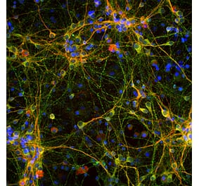
![Immunofluorescence - Anti-Tau Antibody [2E9] (A85416) - Antibodies.com](https://cdn.antibodies.com/image/catalog/85/A85416_1.jpg?profile=product_alternative)
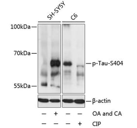
![Western Blot - Anti-Tau (phospho Thr231) Antibody [ARC0021] (A309486) - Antibodies.com](https://cdn.antibodies.com/image/catalog/309/A309486_1.jpg?profile=product_alternative)
![Western Blot - Anti-Tau (phospho Ser396) Antibody [ARC1573] (A309489) - Antibodies.com](https://cdn.antibodies.com/image/catalog/309/A309489_1.jpg?profile=product_alternative)
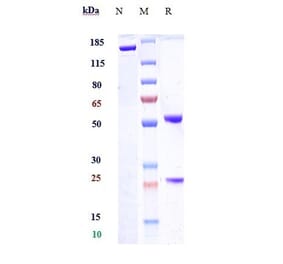
![Immunohistochemistry - Anti-Tau (phospho Ser202 + Thr205) Antibody [AH36] (A304919) - Antibodies.com](https://cdn.antibodies.com/image/catalog/304/A304919_1.png?profile=product_alternative)
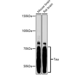
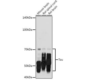
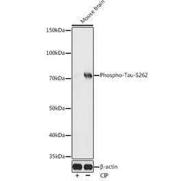
![Western Blot - Anti-Tau Antibody [1D5] (A305068) - Antibodies.com](https://cdn.antibodies.com/image/catalog/305/A305068_1.png?profile=product_alternative)
![Immunocytochemistry/Immunofluorescence - Anti-Tau Antibody [3D4] (A305069) - Antibodies.com](https://cdn.antibodies.com/image/catalog/305/A305069_1.png?profile=product_alternative)
