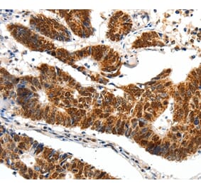
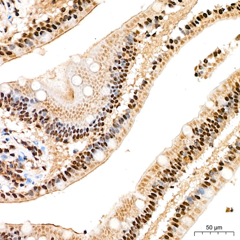
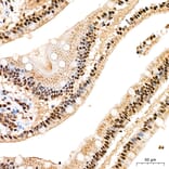
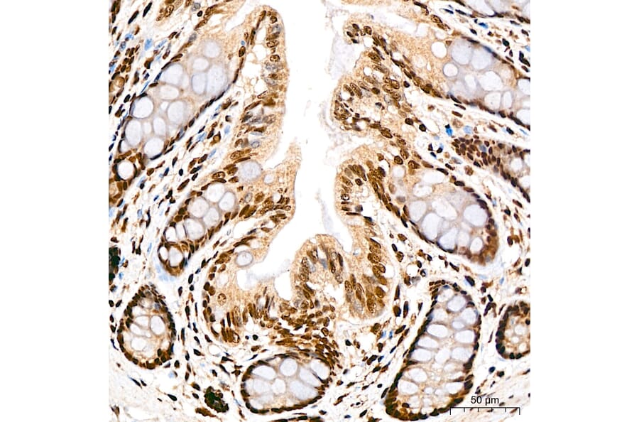
SP1 expression in human small intestine tissue analyzed by immunohistochemistry. Tissue was paraffin-embedded, and antigen retrieval was achieved with 10 mM citrate buffer, pH 6.0, under high pressure. Staining was performed with Anti-SP1 Antibody (A329868) at a dilution of 1:200.
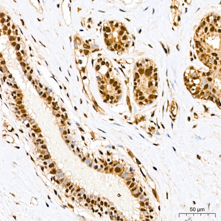
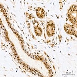
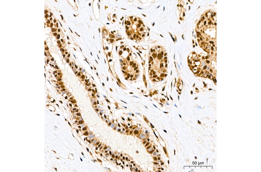
SP1 expression in human breast tissue analyzed by immunohistochemistry. Tissue was paraffin-embedded, and antigen retrieval was achieved with 10 mM citrate buffer, pH 6.0, under high pressure. Staining was performed with Anti-SP1 Antibody (A329868) at a dilution of 1:200.
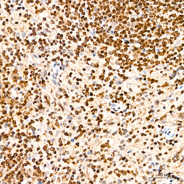
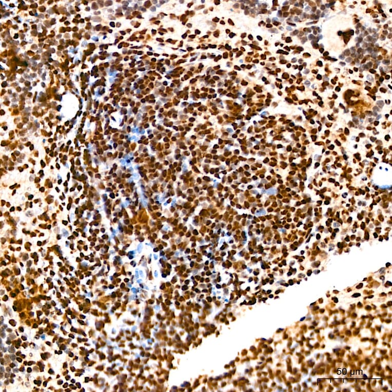
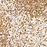
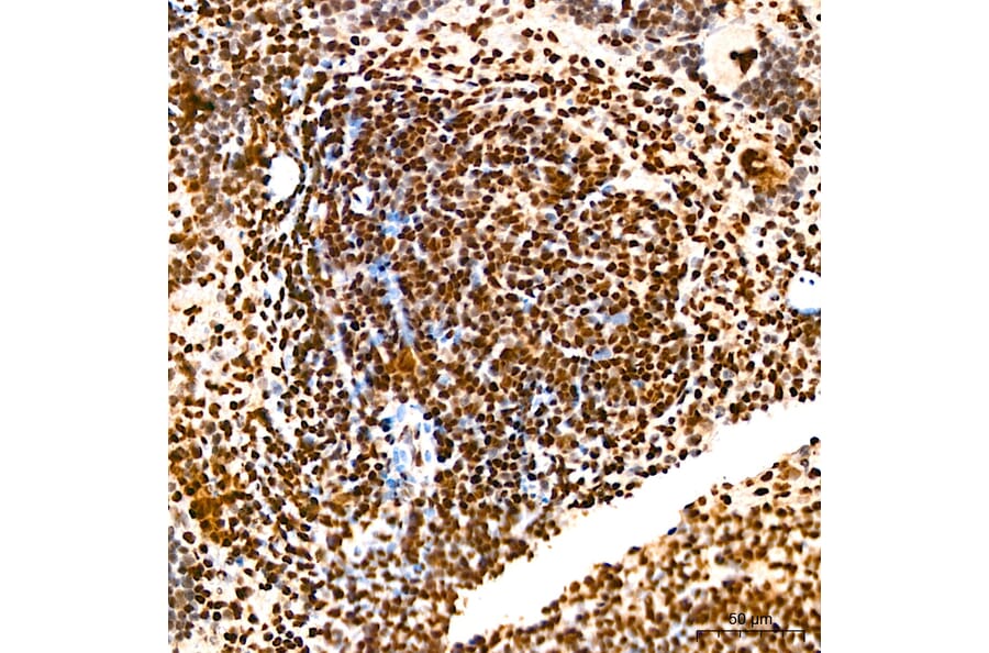
SP1 expression in human spleen tissue analyzed by immunohistochemistry. Tissue was paraffin-embedded, and antigen retrieval was achieved with 10 mM citrate buffer, pH 6.0, under high pressure. Staining was performed with Anti-SP1 Antibody (A329868) at a dilution of 1:200.
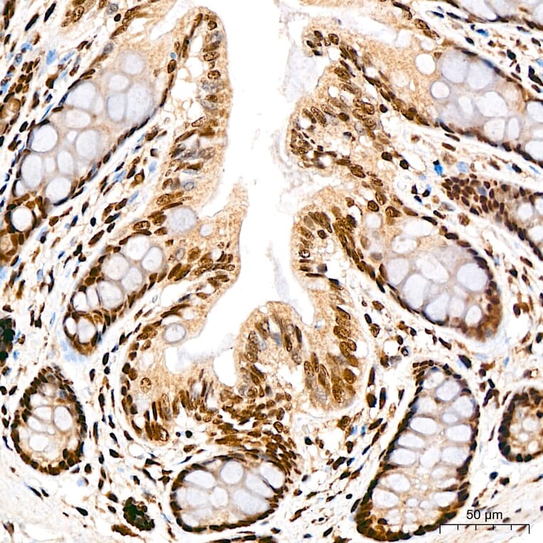
SP1 expression in rat colon tissue analyzed by immunohistochemistry. Tissue was paraffin-embedded, and antigen retrieval was achieved with 10 mM citrate buffer, pH 6.0, under high pressure. Staining was performed with Anti-SP1 Antibody (A329868) at a dilution of 1:200.
SP1 expression in mouse spleen tissue analyzed by immunohistochemistry. Tissue was paraffin-embedded, and antigen retrieval was achieved with 10 mM citrate buffer, pH 6.0, under high pressure. Staining was performed with Anti-SP1 Antibody (A329868) at a dilution of 1:200.


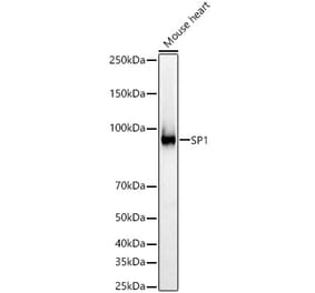
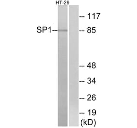
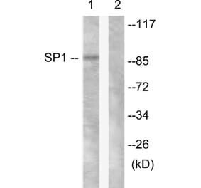
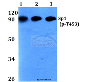
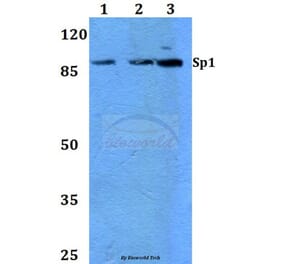
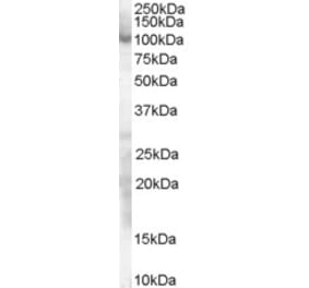
![Western Blot - Anti-SP1 Antibody [ARC0128] (A308986) - Antibodies.com](https://cdn.antibodies.com/image/catalog/308/A308986_1.jpg?profile=product_alternative)