Supplied in Phosphate Buffered Saline, pH 7.3, with 50% Glycerol and 0.02% Sodium Azide.
Storage
Shipped at 4°C. Upon delivery aliquot and store at -20°C. Avoid freeze/thaw cycles.
Synonyms
NAD-dependent protein deacetylase sirtuin-2, NAD-dependent protein defatty-acylase sirtuin-2, Regulatory protein SIR2 homolog 2, SIR2-like protein 2, SIR2L, SIR2L2
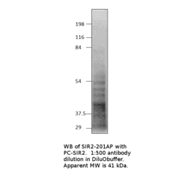
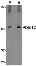
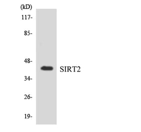
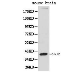
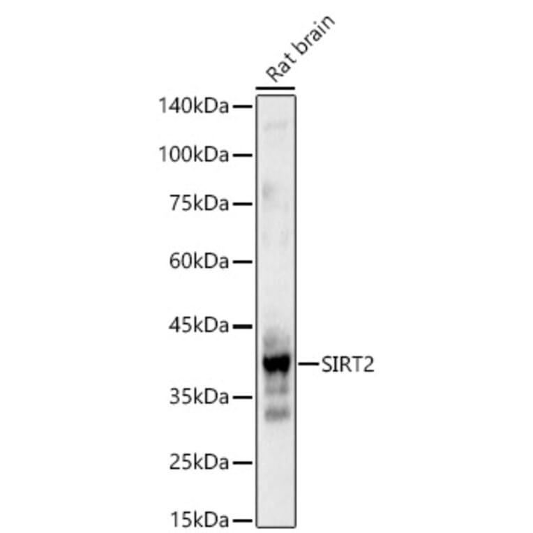
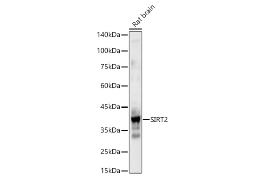
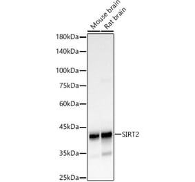
Western blot analysis of Rat brain, using Anti-SIRT2 Antibody (A87642) at 1:1,000 dilution. The secondary antibody was Goat Anti-Rabbit IgG H&L Antibody (HRP) at 1:10,000 dilution. Lysates/proteins were present at 25µg per lane. The blocking buffer used was 3% non-fat dry milk in TBST. Detection was with a ECL Basic Kit. Exposure time: 0. 8s.
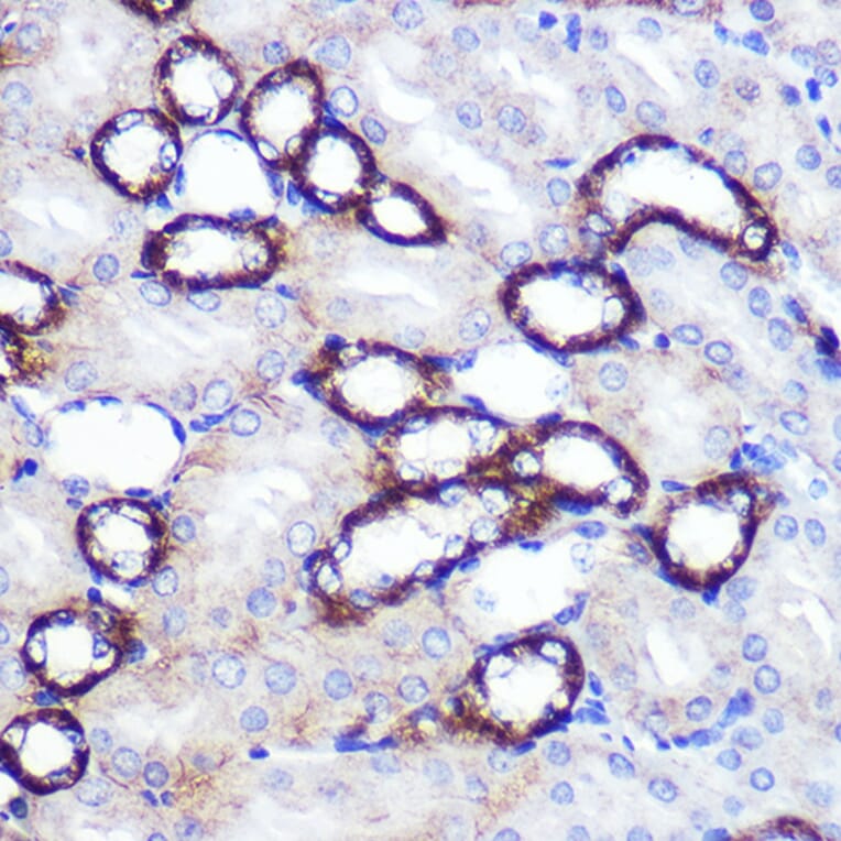
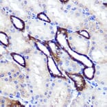
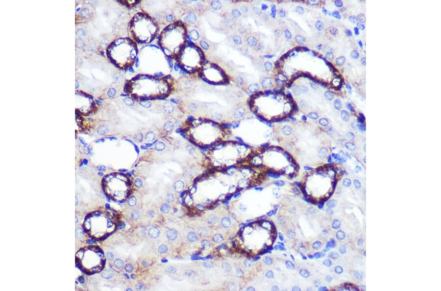
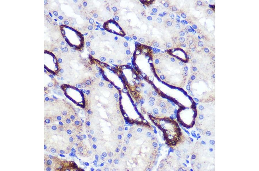
Immunohistochemistry analysis of paraffin-embedded mouse kidney using Anti-SIRT2 Antibody (A87642) at a dilution of 1:100 (40x lens). Perform microwave antigen retrieval with 10 mM PBS buffer pH 7.2 before commencing with IHC staining protocol.
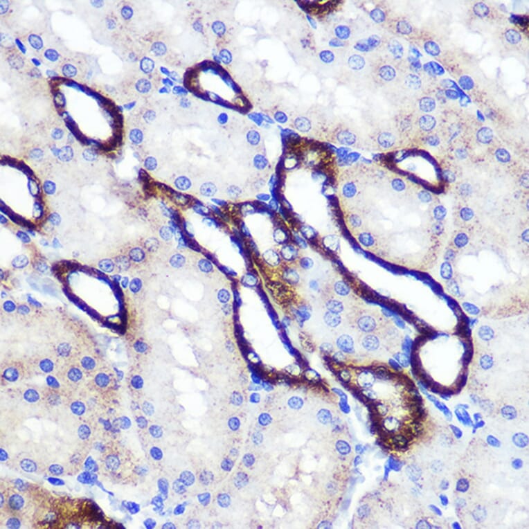
Immunohistochemistry analysis of paraffin-embedded rat kidney using Anti-SIRT2 Antibody (A87642) at a dilution of 1:100 (40x lens). Perform microwave antigen retrieval with 10 mM PBS buffer pH 7.2 before commencing with IHC staining protocol.
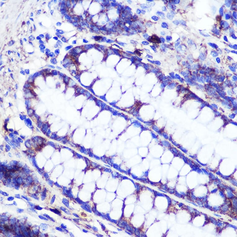
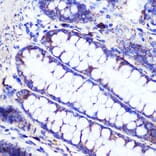
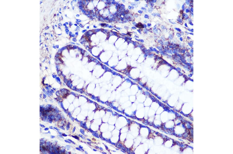
Immunohistochemistry analysis of paraffin-embedded human colon tissue using Anti-SIRT2 Antibody (A87642) at a dilution of 1:100 (40x lens). Perform microwave antigen retrieval with 10 mM PBS buffer pH 7.2 before commencing with IHC staining protocol.
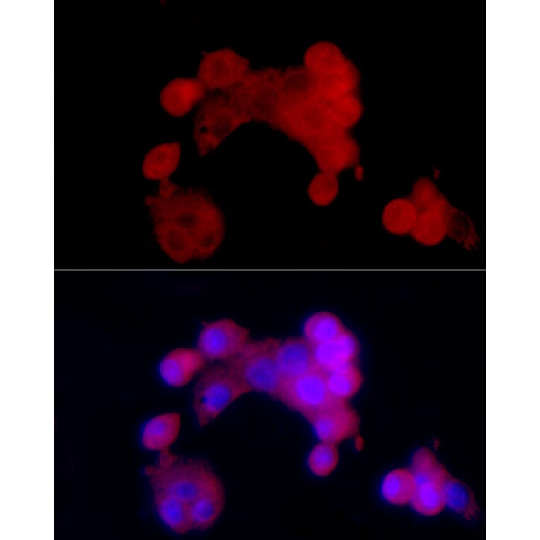
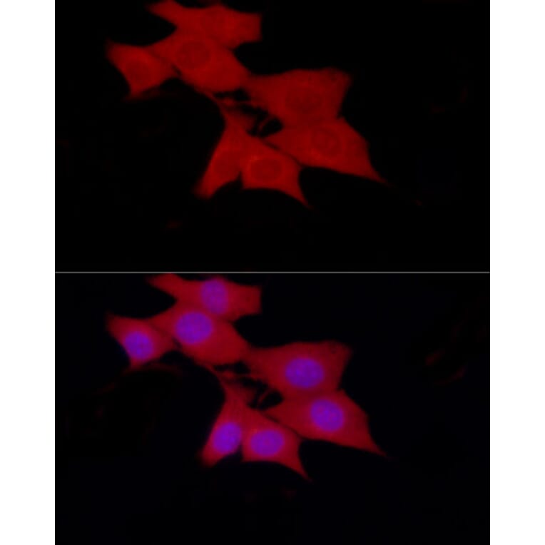
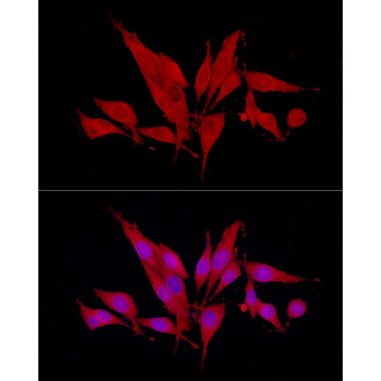
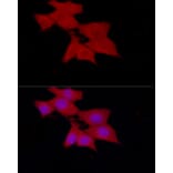
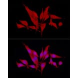
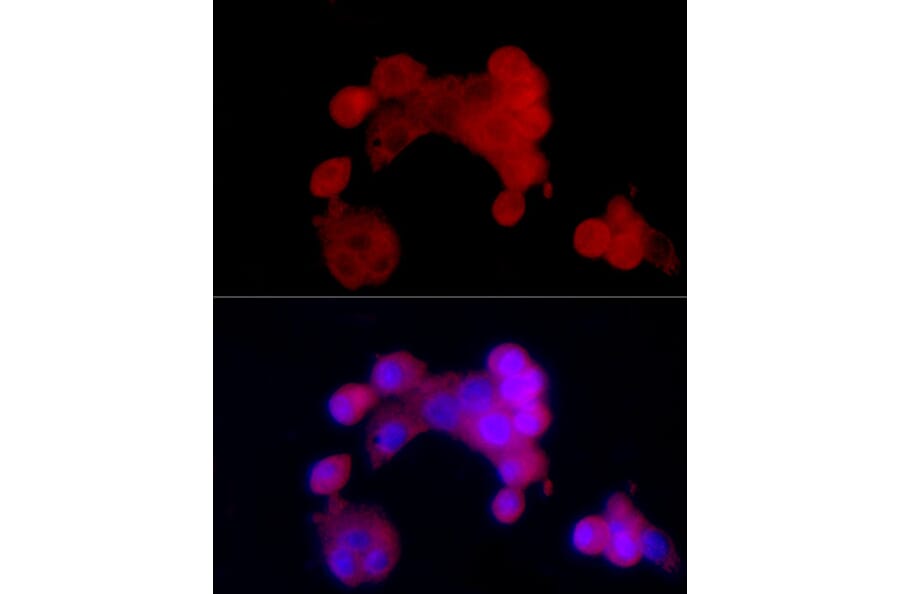
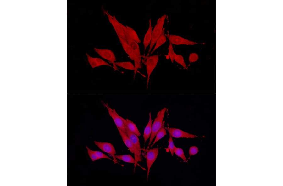
Immunofluorescence analysis of HepG2 cells using Anti-SIRT2 Antibody (A87642) at a dilution of 1:100 (40x lens). DAPI was used to stain the cell nuclei (blue).
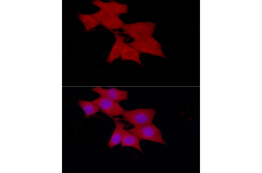
Immunofluorescence analysis of NIH/3T3 cells using Anti-SIRT2 Antibody (A87642) at a dilution of 1:100 (40x lens). DAPI was used to stain the cell nuclei (blue).
Immunofluorescence analysis of PC-12 cells using Anti-SIRT2 Antibody (A87642) at a dilution of 1:100 (40x lens). DAPI was used to stain the cell nuclei (blue).


![Flow Cytometry - Anti-SIRT2 Antibody [PCRP-SIRT2-1A8] (A248540) - Antibodies.com](https://cdn.antibodies.com/image/catalog/248/A248540_1.jpg?profile=product_alternative)
![Flow Cytometry - Anti-SIRT2 Antibody [PCRP-SIRT2-1A8] - BSA and Azide free (A251722) - Antibodies.com](https://cdn.antibodies.com/image/catalog/251/A251722_1.jpg?profile=product_alternative)