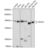
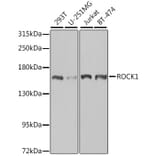
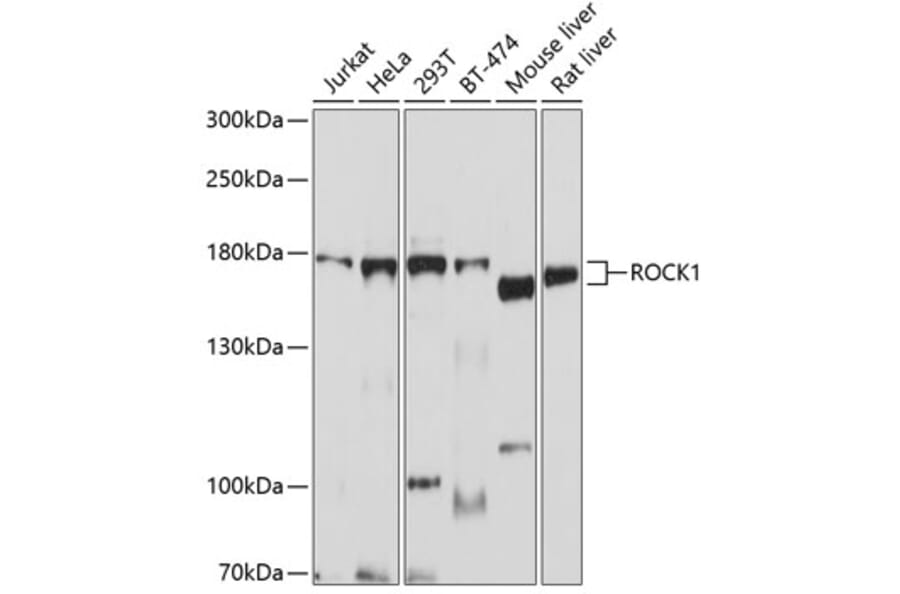
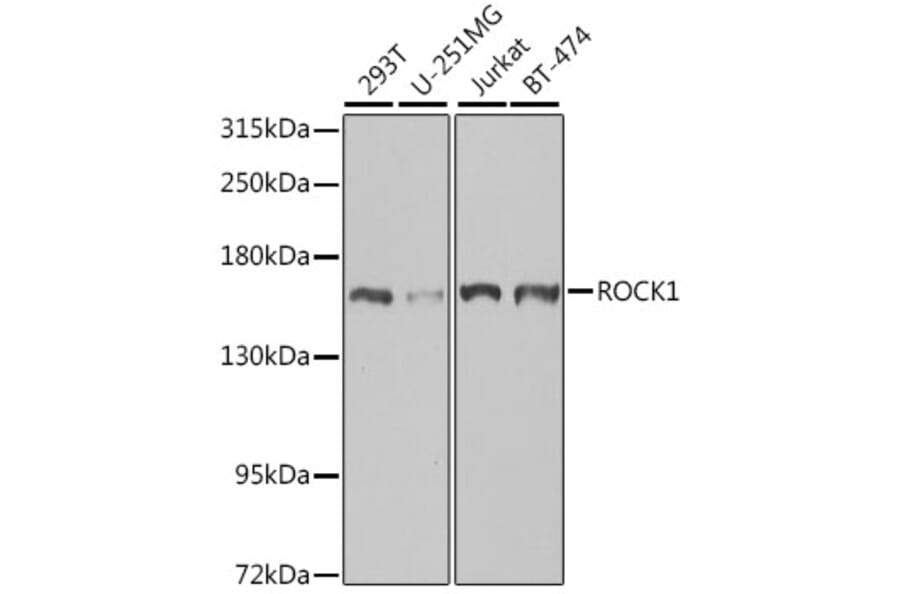
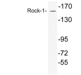
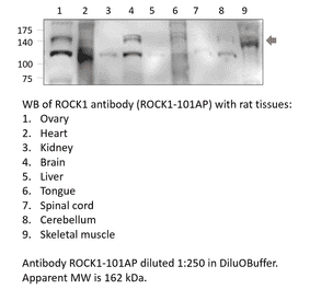
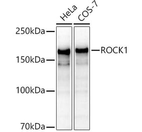
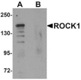
ROCK1 expression in various lysates analyzed by western blot. Primary antibody incubation was performed for 1 hour on 25ug protein per lane with Anti-ROCK1 Antibody (A12527) at a dilution of 1:1000 and detected with chemiluminescence.
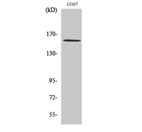
ROCK1 expression in various lysates analyzed by western blot. Primary antibody incubation was performed for 1 hour on 25ug protein per lane with Anti-ROCK1 Antibody (A12527) at a dilution of 1:1000 and detected with chemiluminescence.
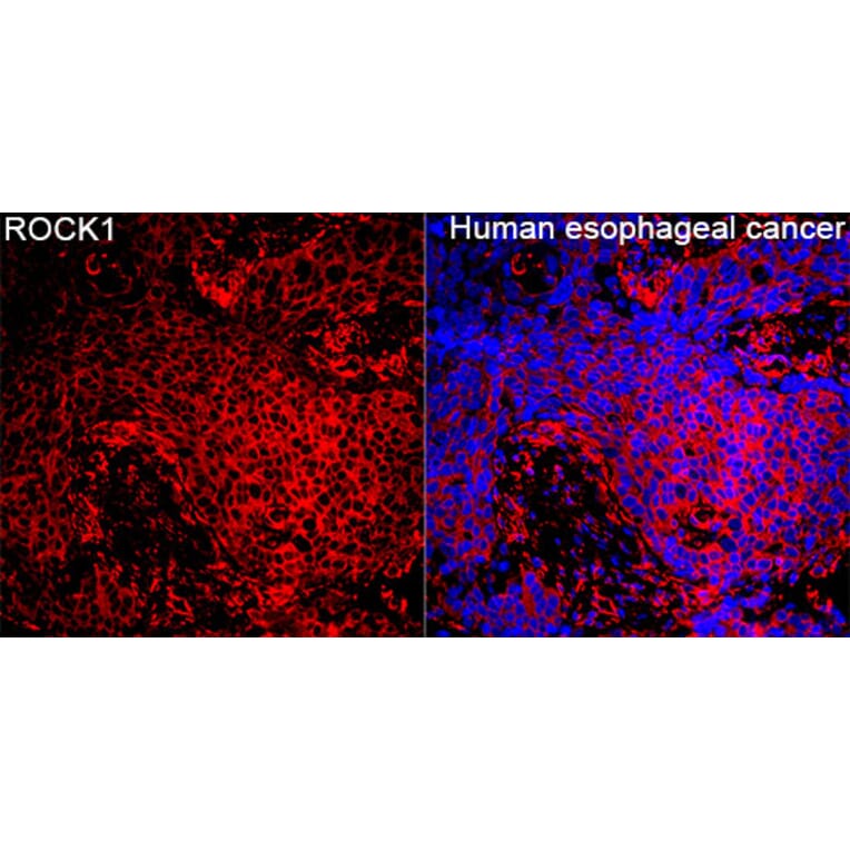
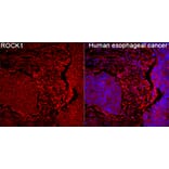
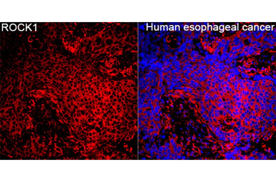
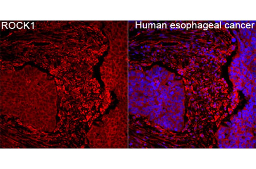
ROCK1 expression in Human esophageal cancer tissue analyzed by immunofluorescence. Staining was performed with Anti-ROCK1 Antibody (A12527) at a dilution of 1:100 followed by Cy3-conjugated Goat anti-Rabbit IgG (H+L)secondary antibody at a dilution of 1:500. Nuclei were stained with DAPI (blue).
ROCK1 expression in Human esophageal cancer tissue analyzed by immunofluorescence. Staining was performed with Anti-ROCK1 Antibody (A12527) at a dilution of 1:100 followed by Cy3-conjugated Goat anti-Rabbit IgG (H+L)secondary antibody at a dilution of 1:500. Nuclei were stained with DAPI (blue).
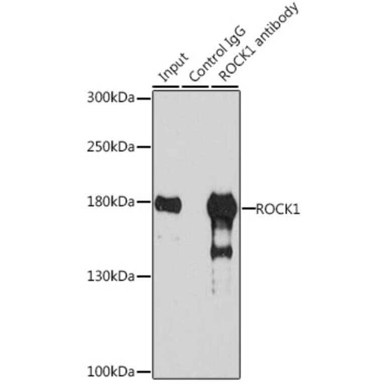
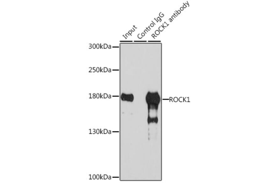
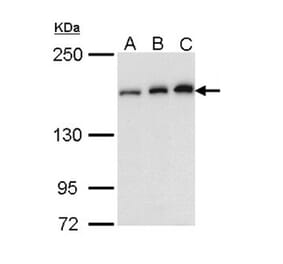
ROCK1 expression in 200 µg extracts of 293T cells analyzed by western blot following immunoprecipitation, both using Anti-ROCK1 Antibody (A12527) at 3 µg and a dilution of 1:1000, respectively.


![Western Blot - Anti-ROCK1 Antibody [ARC2371] (A13244) - Antibodies.com](https://cdn.antibodies.com/image/catalog/13/A13244_1.jpg?profile=product_alternative)