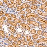
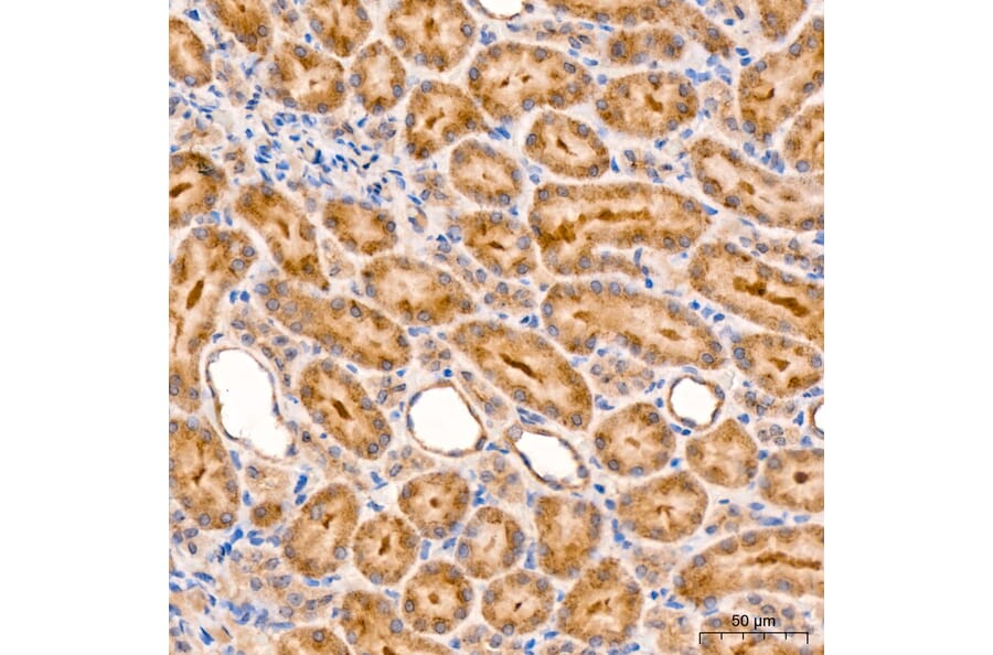
RIP3 expression in rat kidney tissue analyzed by immunohistochemistry. Tissue was paraffin-embedded, and antigen retrieval was achieved with 10 mM citrate buffer, pH 6.0, under high pressure. Staining was performed with Anti-RIP3 Antibody (A329805) at a dilution of 1:100.
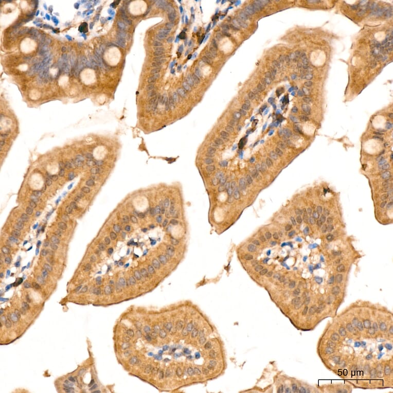
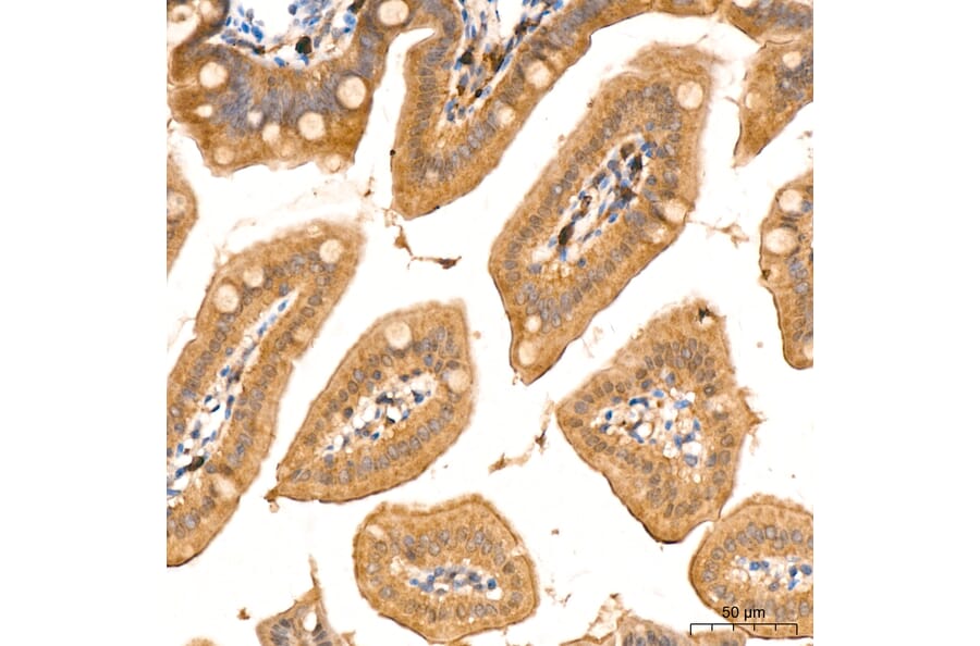
RIP3 expression in mouse intestin tissue analyzed by immunohistochemistry. Tissue was paraffin-embedded, and antigen retrieval was achieved with 10 mM citrate buffer, pH 6.0, under high pressure. Staining was performed with Anti-RIP3 Antibody (A329805) at a dilution of 1:100.
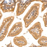
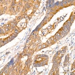
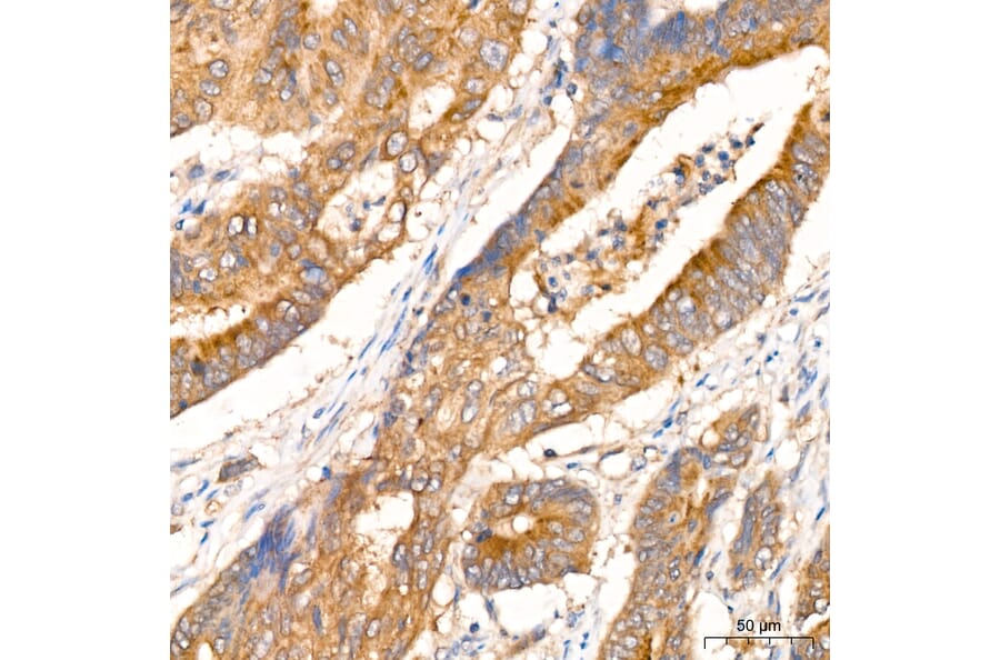
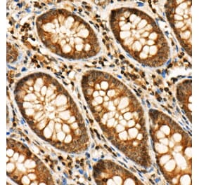
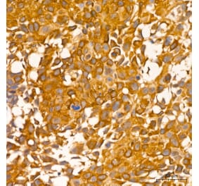
RIP3 expression in human colon carcinoma tissue analyzed by immunohistochemistry. Tissue was paraffin-embedded, and antigen retrieval was achieved with 10 mM citrate buffer, pH 6.0, under high pressure. Staining was performed with Anti-RIP3 Antibody (A329805) at a dilution of 1:100.
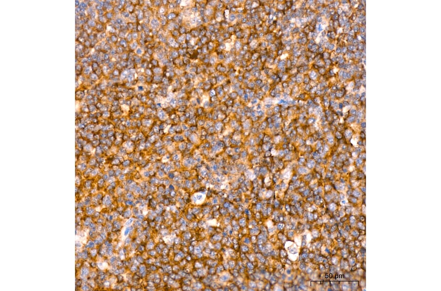
RIP3 expression in human tonsil tissue analyzed by immunohistochemistry. Tissue was paraffin-embedded, and antigen retrieval was achieved with 10 mM citrate buffer, pH 6.0, under high pressure. Staining was performed with Anti-RIP3 Antibody (A329805) at a dilution of 1:100.
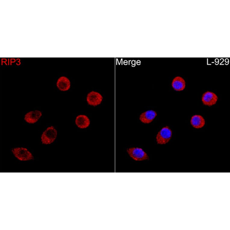
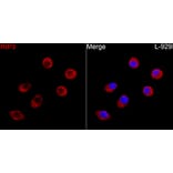
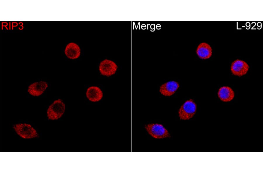
RIP3 expression in L929 cells analyzed by immunofluorescence. Staining was performed with Anti-RIP3 Antibody (A329805) at a dilution of 1:100 followed by Cy3 Goat Anti-Rabbit IgG (H+L) secondary antibody at a dilution of 1:500. Nuclei were stained with DAPI (blue).