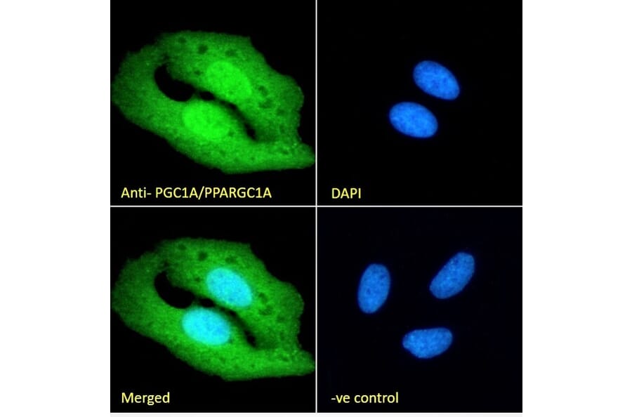
PGC1 alpha expression in U2OS cells analyzed by immunofluorescence. Cells were permeabilized with 0.15% Triton. Staining was performed with Anti-PGC1 alpha Antibody (A84356) at 10µg/ml for 1 hour and Alexa Fluor 488 secondary antibody at 2µg/ml. Cytoplasmic and nuclear staining shown and nuclei were stained with DAPI (blue). Negative control: Goat IgG Isotype Control at 10µg/ml followed by Alexa Fluor 488 secondary antibody at 2µg/ml.
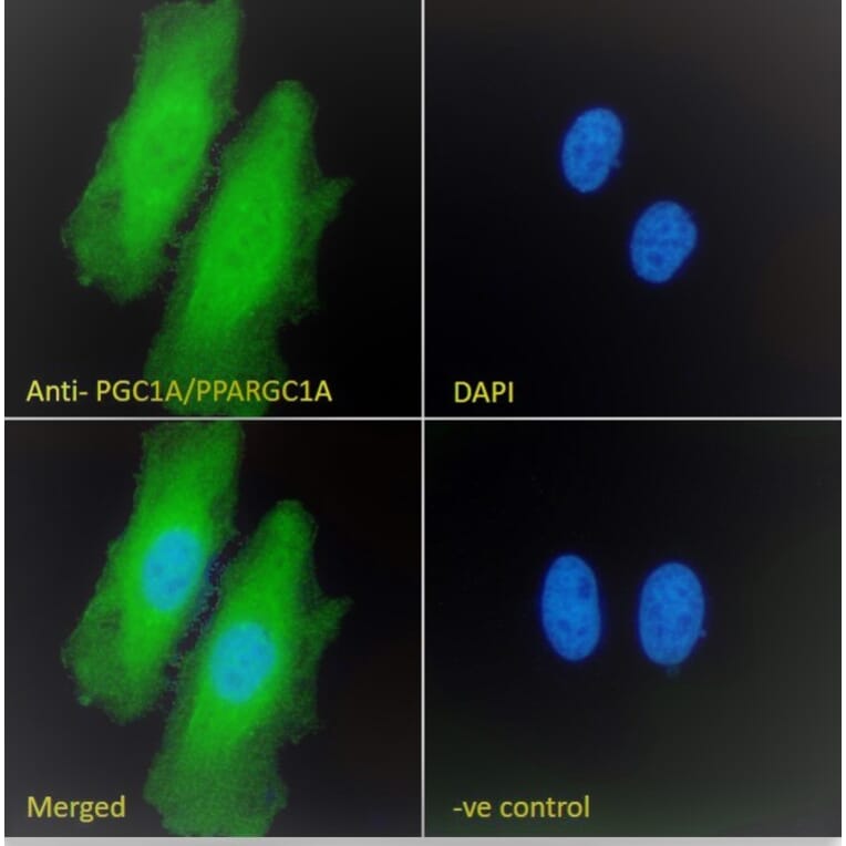
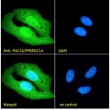
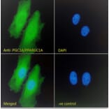
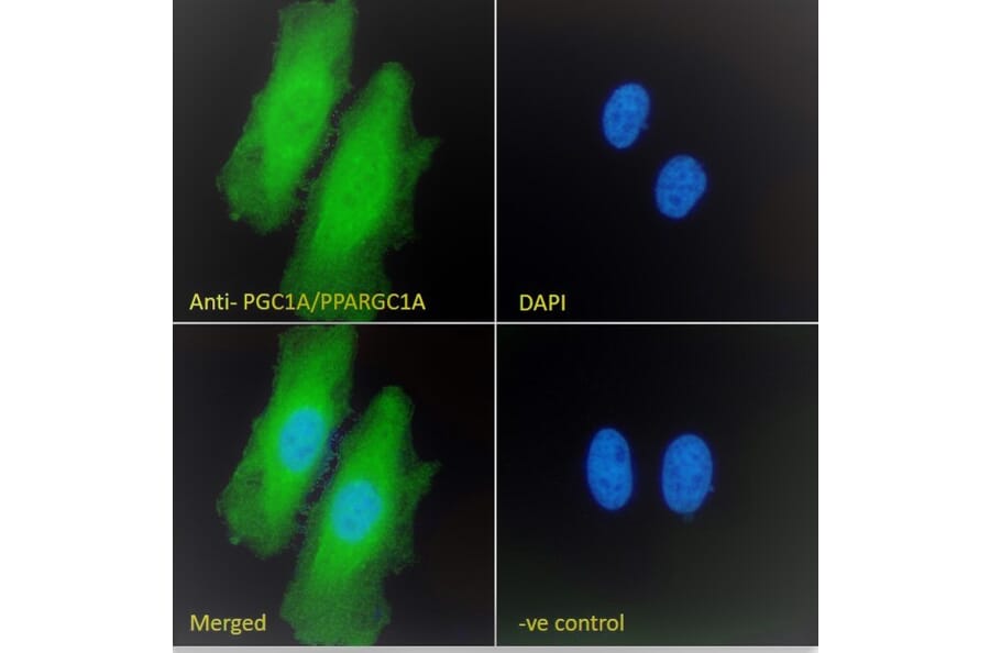
PGC1 alpha expression in HeLa cells analyzed by immunofluorescence. Cells were permeabilized with 0.15% Triton. Staining was performed with Anti-PGC1 alpha Antibody (A84356) at 10µg/ml for 1 hour and Alexa Fluor 488 secondary antibody at 2µg/ml. Cytoplasmic and nuclear staining shown and nuclei were stained with DAPI (blue). Negative control: Goat IgG Isotype Control at 10µg/ml followed by Alexa Fluor 488 secondary antibody at 2µg/ml.
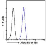
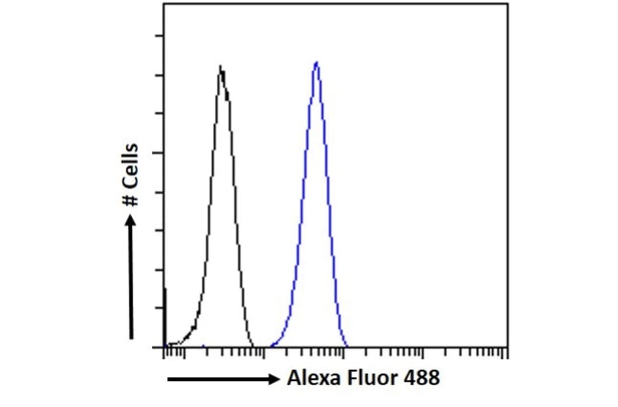
PGC1 alpha expression in HeLa cells (blue line) analyzed by flow cytometry. Cells were fixed in PFA and permeabilized with 0.5% Triton. Staining was performed with Anti-PGC1 alpha Antibody (A84356) at 10µg/ml for 1 hour and Alexa Fluor 488 secondary antibody at 1µg/ml. Negative Control: Goat IgG Isotype Control (black line) followed by Alexa Fluor 488 secondary antibody.
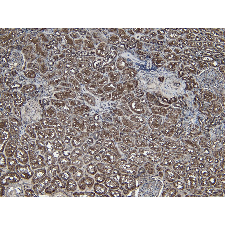
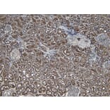
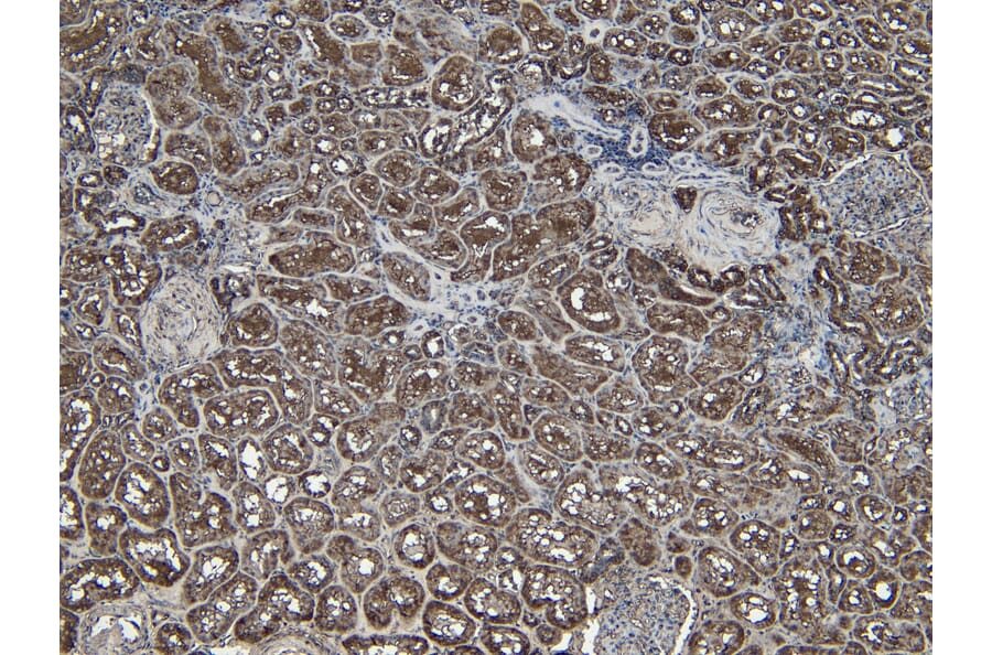
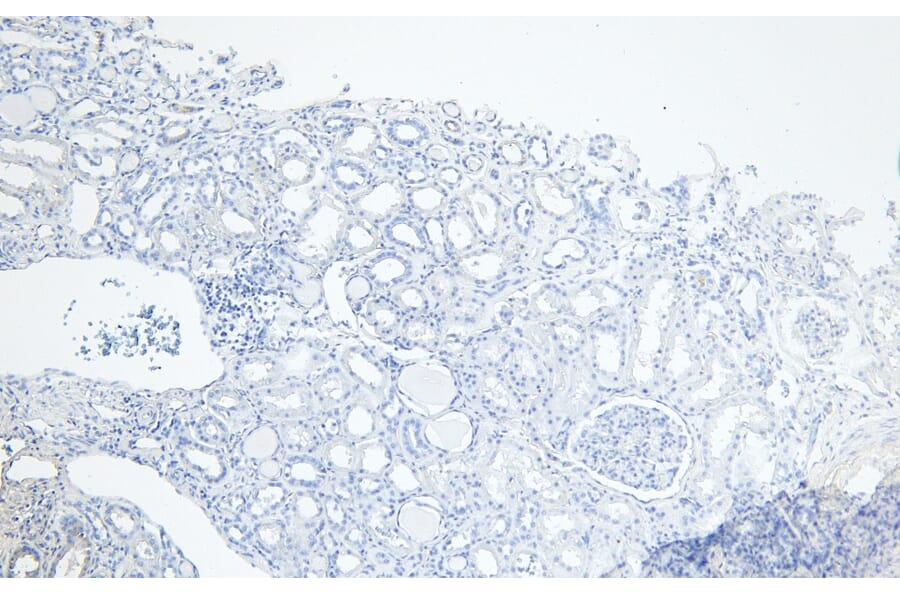
PGC1 alpha expression in Human Kidney analyzed by immunohistochemistry. Tissue was paraffin-embedded, and antigen retrieval was achieved by heating in citrate buffer, pH 6. Staining was performed with Anti-PGC1 alpha Antibody (A84356) at 6µg/ml and revealed with horseradish peroxidase (HRP).
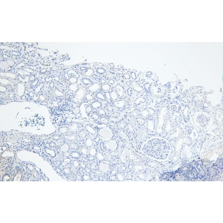
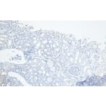
Negative control for PGC1 alpha expression in Human Kidney analyzed by immunohistochemistry. Tissue was paraffin-embedded, and staining procedure was performed in the absence of primary antibody.