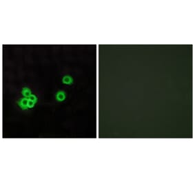
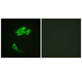
Immunofluorescent analysis of rat brain section stained with Anti-PEA15 Antibody [4D117] (A270552), at a dilution of 1:1,000, in red, and co-stained with Anti-GFAP Antibody (A85419), at a dilution of 1:5,000, in green. Nuclear DNA is visualised in blue using Hoechst staining. Following transcardial perfusion with 4% paraformaldehyde, the brain was post-fixed for 24 hours, cut to 45 µm, and free-floating sections were stained using the above antibodies. Anti-PEA15 Antibody [4D117] (A270552) labels the cytoplasm of certain presumably neuronal cells which are not labelled by the astrocyte specific Anti-GFAP Antibody (A85419).
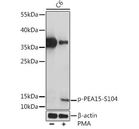
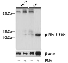
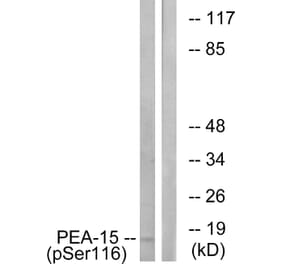
Western Blot - Anti-PEA15 Antibody [4D117] (A270552)
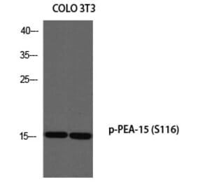
Western blot analysis of different tissue lysates using Anti-PEA15 Antibody [4D117] (A270552), at a dilution of 1:1,000, in green. The lanes contain: [Lane 1] protein standard (red(, [Lane 2] rat whole brain, [Lane 3] rat cerebellum, [Lane 4] mouse whole brain, [Lane 5] cow cortex, and [Lane 6] cow cerebellum. The strong band at about 15 kDa corresponds to the PEA15 protein.
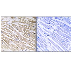
Immunohistochemistry analysis of a 4% PFA fixed paraffin embedded rat testes section with Anti-PEA15 Antibody [4D117] (A270552) at a dilution of 1:1,000 detected with DAB (brown) using the Vector Labs ImmPRESS method and reagents with citra buffer retrieval. Counterstained with Hematoxylin (blue). Note: this antibody performs well in testing with 4% PFA but does not stain long term NBF fixed tissue effectively.
Publishing research using Anti-PEA15 Antibody [4D117] (A270552)? Please let us know so that we can list the citation on this page.
Alternative products to Anti-PEA15 Antibody [4D117] (A270552)


![Immunofluorescence - Anti-PEA15 Antibody [4D117] (A270552) - Antibodies.com](https://cdn.antibodies.com/image/catalog/270/A270552_1.jpg?profile=product_top)
![Western Blot - Anti-PEA15 Antibody [4D117] (A270552) - Antibodies.com](https://cdn.antibodies.com/image/catalog/270/A270552_2.jpg?profile=product_top)
![Immunohistochemistry - Anti-PEA15 Antibody [4D117] (A270552) - Antibodies.com](https://cdn.antibodies.com/image/catalog/270/A270552_3.jpg?profile=product_top)
![Immunofluorescence - Anti-PEA15 Antibody [4D117] (A270552) - Antibodies.com](https://cdn.antibodies.com/image/catalog/270/A270552_1.jpg?profile=product_top_thumb)
![Western Blot - Anti-PEA15 Antibody [4D117] (A270552) - Antibodies.com](https://cdn.antibodies.com/image/catalog/270/A270552_2.jpg?profile=product_top_thumb)
![Immunohistochemistry - Anti-PEA15 Antibody [4D117] (A270552) - Antibodies.com](https://cdn.antibodies.com/image/catalog/270/A270552_3.jpg?profile=product_top_thumb)
![Immunofluorescence - Anti-PEA15 Antibody [4D117] (A270552) - Antibodies.com](https://cdn.antibodies.com/image/catalog/270/A270552_1.jpg?profile=product_image)
![Western Blot - Anti-PEA15 Antibody [4D117] (A270552) - Antibodies.com](https://cdn.antibodies.com/image/catalog/270/A270552_2.jpg?profile=product_image)
![Immunohistochemistry - Anti-PEA15 Antibody [4D117] (A270552) - Antibodies.com](https://cdn.antibodies.com/image/catalog/270/A270552_3.jpg?profile=product_image)
![Immunofluorescence - Anti-PEA15 Antibody [4D2] (A270553) - Antibodies.com](https://cdn.antibodies.com/image/catalog/270/A270553_1.jpg?profile=product_alternative)