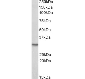
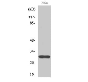
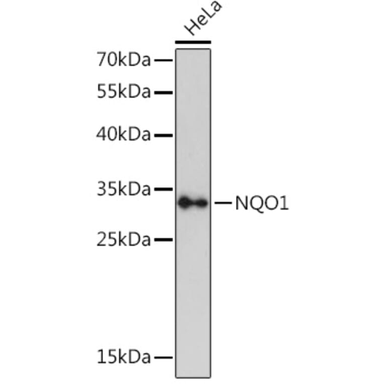
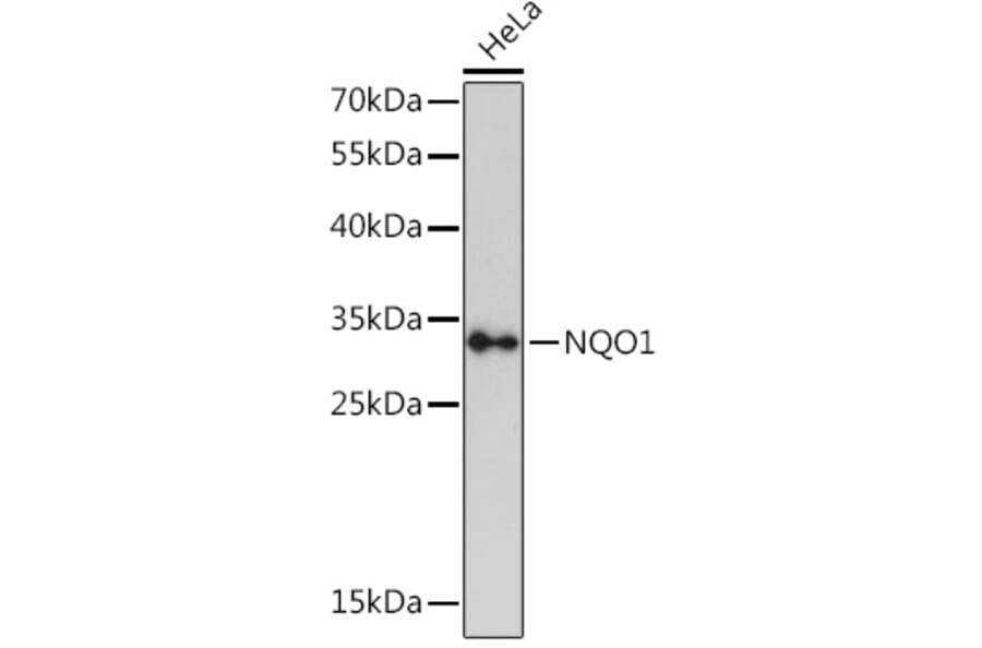
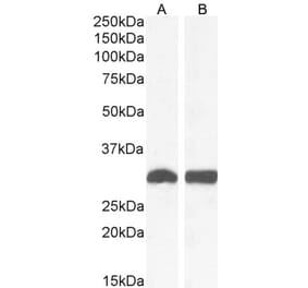
NQO1 expression in HeLa cell lysates analyzed by western blot. Primary antibody incubation was performed for 1 hour on 25ug protein per lane with Anti-NQO1 Antibody (A12506) at a dilution of 1:3000 and detected with chemiluminescence.
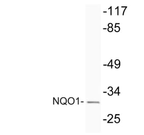
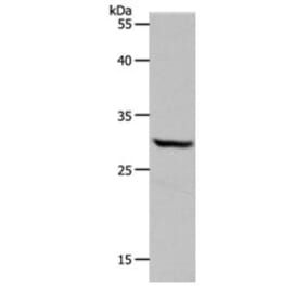
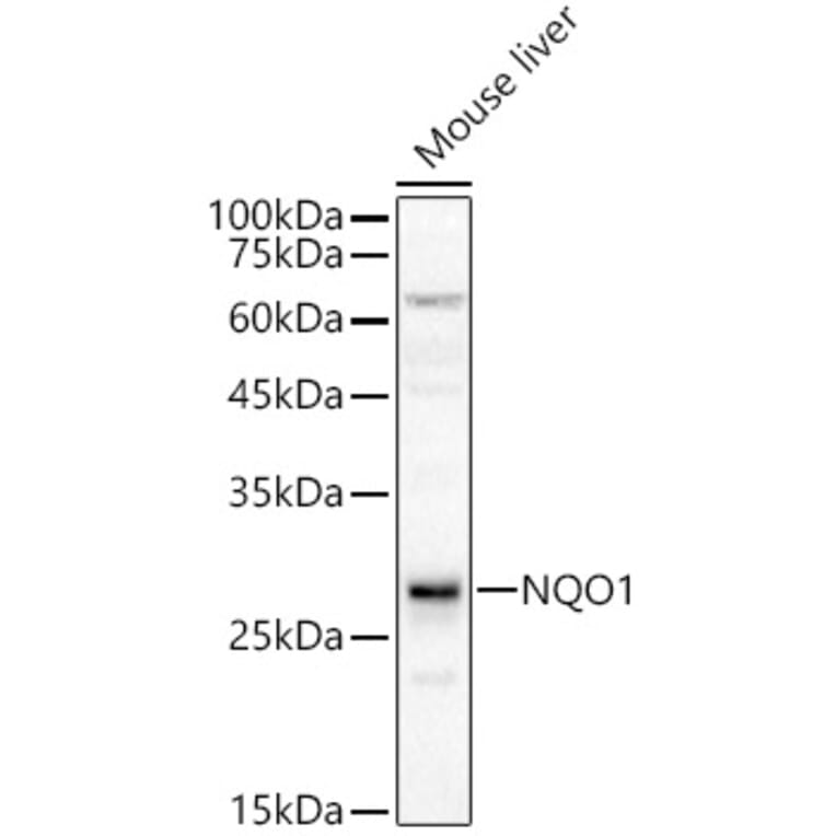
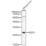
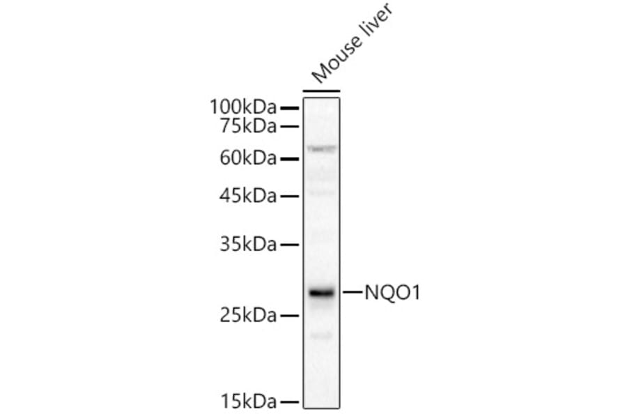
NQO1 expression in Mouse liver lysates analyzed by western blot. Primary antibody incubation was performed for 1 hour on 25ug protein per lane with Anti-NQO1 Antibody (A12506) at a dilution of 1:400 and detected with chemiluminescence.
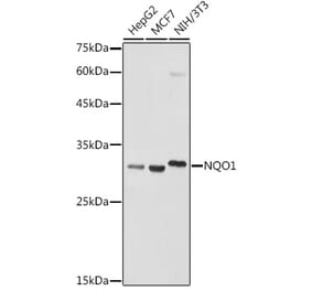
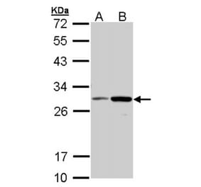
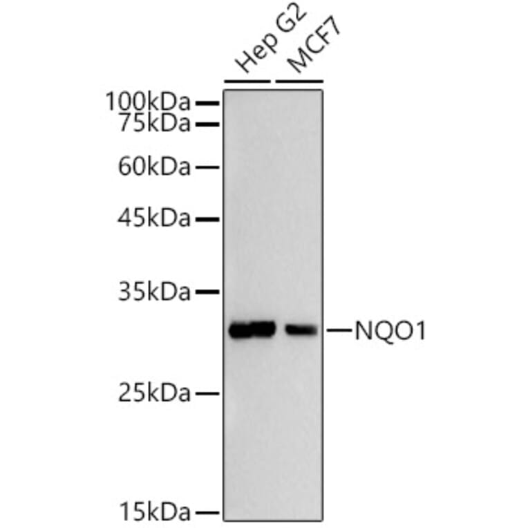
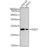
NQO1 expression in various lysates analyzed by western blot. Primary antibody incubation was performed for 1 hour on 25ug protein per lane with Anti-NQO1 Antibody (A12506) at a dilution of 1:500 and detected with chemiluminescence.
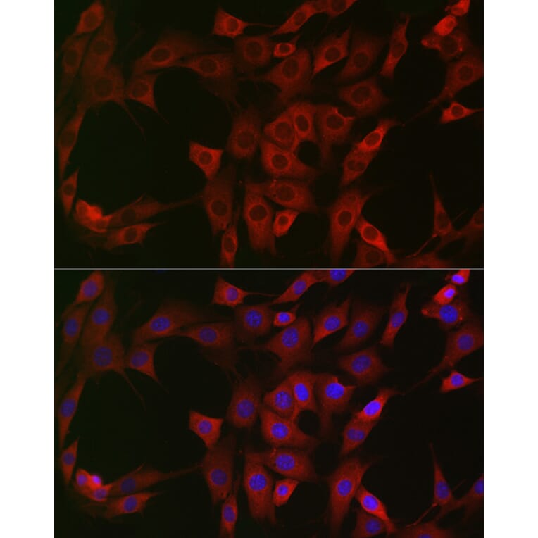
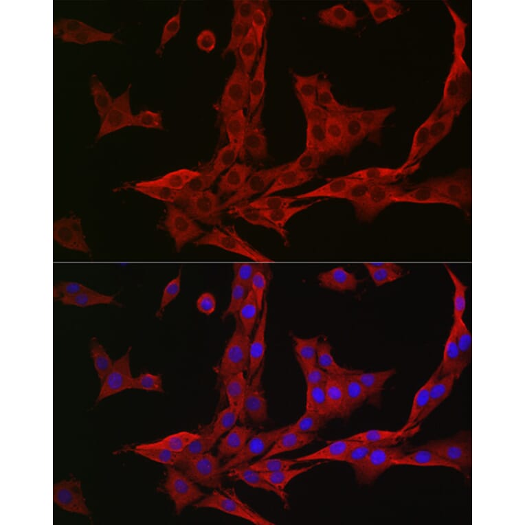
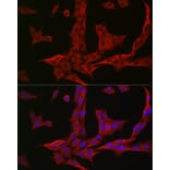
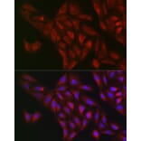
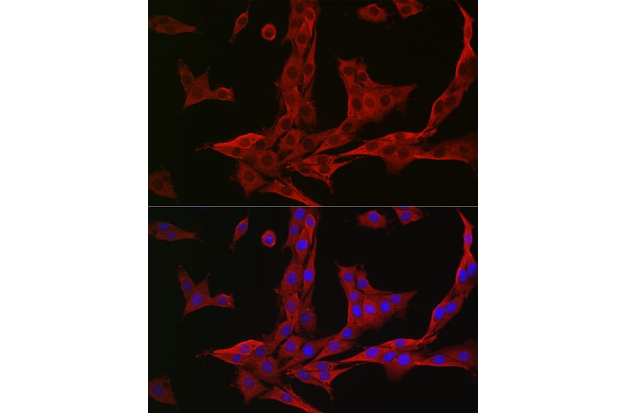
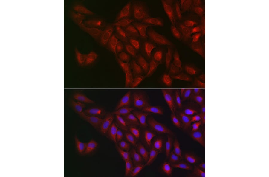
NQO1 expression in NIH/3T3 cells analyzed by immunofluorescence. Staining was performed with Anti-NQO1 Antibody (A12506) at a dilution of 1:50 followed by Cy3-conjugated Goat anti-Rabbit IgG (H+L) secondary antibody at a dilution of 1:500. Nuclei were stained with DAPI (blue).
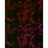
NQO1 expression in PC-12 cells analyzed by immunofluorescence. Staining was performed with Anti-NQO1 Antibody (A12506) at a dilution of 1:50 followed by Cy3-conjugated Goat anti-Rabbit IgG (H+L) secondary antibody at a dilution of 1:500. Nuclei were stained with DAPI (blue).
NQO1 expression in U2OS cells analyzed by immunofluorescence. Staining was performed with Anti-NQO1 Antibody (A12506) at a dilution of 1:50 followed by Cy3-conjugated Goat anti-Rabbit IgG (H+L) secondary antibody at a dilution of 1:500. Nuclei were stained with DAPI (blue).


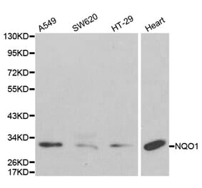
![Western Blot - Anti-NQO1 Antibody [ARC56753] (A307961) - Antibodies.com](https://cdn.antibodies.com/image/catalog/307/A307961_1.jpg?profile=product_alternative)