This antibody recognizes phosphorylated NF-H KSP sequences but not non-phosphorylated KSP sequences. In some species there is cross-reactivity with the phosphorylated KSP sequences found in the related neurofilament subunit NF-M.
Applications
WB, ICC/IF, IHC
Dilutions
WB: 1:10,000, ICC/IF: 1:1,000, IHC: 1:4,000
Reactivity
Human, Rat, Mouse, Bovine, Porcine, Horse
Immunogen
Native axonal phosphorylated NF-H, purified from bovine spinal cord.
Host
Mouse
Clonality
Monoclonal
Clone ID
AH1
Isotype
IgG1
Light Chains
kappa
Conjugate
Unconjugated
Purification
Immunogen affinity purification.
Concentration
1 mg/ml
Molecular Weight
200-220 kDa
Product Form
Liquid
Formulation
Supplied in Phosphate Buffered Saline with 50% Glycerol and 5mM Sodium Azide.
Storage
Shipped at 4°C. Upon delivery aliquot and store at -20°C. Avoid freeze/thaw cycles.
Synonyms
200 kDa neurofilament protein, KIAA0845, NEFH, Neurofilament heavy polypeptide, Neurofilament triplet H protein, NFH
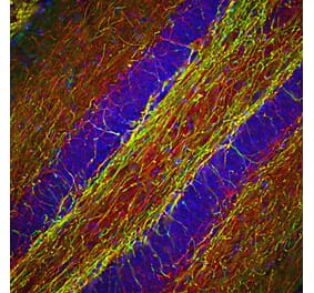
Immunofluorescent analysis of rat brain section stained with Anti-Tyrosine Hydroxylase Antibody (A104319), at a dilution of 1:10,000, in red, and co-stained with Anti-NF-H Antibody [AH1] (A85340), at a dilution of 1:1,000, in green. The blue is Hoechst staining of nuclear DNA. Following transcardial perfusion of rat with 4% paraformaldehyde, brain was post fixed for 24 hours, cut to 45µM, and free-floating sections were stained with the above antibodies. Anti-Tyrosine Hydroxylase Antibody (A104319) stains the striatal TH expressing interneurons, while Anti-NF-H Antibody [AH1] (A85340) labels axons from other neuronal cells.
Western blot detection of the heavily phosphorylated axonal form of NF-H protein (pNF-H) in neural tissue lysates (20µg/lane) with Anti-Neurofilament Heavy Polypeptide Antibody [AH1] (A85340) at dilution of 1:5,000. Lanes on the blot are: [Lane 1] Protein size marker, [Lane 2] Adult rat whole brain [Lane 3] Embryonic (E20) rat whole brain [Lane 4] Adult rat spinal cord [Lane 5] Adult mouse whole brain [Lane 6] Adult mouse spinal cord. Rodent pNF-H protein appears as a single band of about 200 kDa in adult rat and mouse lysates, but is not present in early development (Lane 3). Additional bands appearing on the blot (Lane 4) are most likely partially degraded products of pNF-H protein.
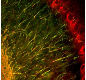
Immunohistochemistry analysis of a formalin fixed paraffin embedded human cerebellum section with Anti-NF-H Antibody [AH1] (A85340) at a dilution of 1:4,000 detected with DAB (brown) using the Vector Labs ImmPRESS method and reagents with citra buffer retrieval. Counterstained with Hematoxylin (blue). Anti-NF-H Antibody [AH1] (A85340) strongly labels basket cell processes and certain parallel fibers and also axons in the molecular and granular layers. Note: this antibody performs well in testing with both 4% PFA and standard NBF fixed rat, mouse and human tissues.
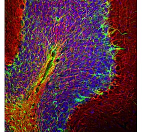
Immunohistological analysis of rat cerebellum section stained with Anti-Neurofilament Heavy Polypeptide Antibody [AH1] (A85340), dilution 1:2,000, in green, and co-stained with Anti-Fox3 Antibody (A85403), dilution 1:5,000, in red. Following transcardial perfusion with 4% paraformaldehyde, the brain was post-fixed for 24 hours, cut to 45 µm, and free-floating sections were stained with the above antibodies. The Anti-Neurofilament Heavy Polypeptide Antibody [AH1] stains axons in the granular layer and white matter and prominent basket cell axons surrounding the large Purkinje neurons. The FOX3 antibody specifically labels nuclei of granular and other neurons, but does not stain Purkinje cells.
Publishing research using Anti-NF-H Antibody [AH1] (A85340)? Please let us know so that we can list the citation on this page.


![Immunofluorescence - Anti-NF-H Antibody [AH1] (A85340) - Antibodies.com](https://cdn.antibodies.com/image/catalog/85/A85340_1.jpg?profile=product_top)
![Western Blot - Anti-NF-H Antibody [AH1] (A85340) - Antibodies.com](https://cdn.antibodies.com/image/catalog/85/A85340_2.jpg?profile=product_top)
![Immunohistochemistry - Anti-NF-H Antibody [AH1] (A85340) - Antibodies.com](https://cdn.antibodies.com/image/catalog/85/A85340_3.jpg?profile=product_top)
![Immunohistochemistry - Anti-NF-H Antibody [AH1] (A85340) - Antibodies.com](https://cdn.antibodies.com/image/catalog/85/A85340_4.jpg?profile=product_top)
![Immunofluorescence - Anti-NF-H Antibody [AH1] (A85340) - Antibodies.com](https://cdn.antibodies.com/image/catalog/85/A85340_1.jpg?profile=product_top_thumb)
![Western Blot - Anti-NF-H Antibody [AH1] (A85340) - Antibodies.com](https://cdn.antibodies.com/image/catalog/85/A85340_2.jpg?profile=product_top_thumb)
![Immunohistochemistry - Anti-NF-H Antibody [AH1] (A85340) - Antibodies.com](https://cdn.antibodies.com/image/catalog/85/A85340_3.jpg?profile=product_top_thumb)
![Immunohistochemistry - Anti-NF-H Antibody [AH1] (A85340) - Antibodies.com](https://cdn.antibodies.com/image/catalog/85/A85340_4.jpg?profile=product_top_thumb)
![Immunofluorescence - Anti-NF-H Antibody [AH1] (A85340) - Antibodies.com](https://cdn.antibodies.com/image/catalog/85/A85340_1.jpg?profile=product_image)
![Western Blot - Anti-NF-H Antibody [AH1] (A85340) - Antibodies.com](https://cdn.antibodies.com/image/catalog/85/A85340_2.jpg?profile=product_image)
![Immunohistochemistry - Anti-NF-H Antibody [AH1] (A85340) - Antibodies.com](https://cdn.antibodies.com/image/catalog/85/A85340_3.jpg?profile=product_image)
![Immunohistochemistry - Anti-NF-H Antibody [AH1] (A85340) - Antibodies.com](https://cdn.antibodies.com/image/catalog/85/A85340_4.jpg?profile=product_image)
![Immunohistochemistry - Anti-Neurofilament Heavy Polypeptide Antibody [NF421] - BSA and Azide free (A252667) - Antibodies.com](https://cdn.antibodies.com/image/catalog/252/A252667_1.jpg?profile=product_alternative)
![Immunohistochemistry - Anti-Neurofilament Heavy Polypeptide Antibody [NE14] (A249491) - Antibodies.com](https://cdn.antibodies.com/image/catalog/249/A249491_1.jpg?profile=product_alternative)
![Immunohistochemistry - Anti-Neurofilament Heavy Polypeptide Antibody [NE14] - BSA and Azide free (A252671) - Antibodies.com](https://cdn.antibodies.com/image/catalog/252/A252671_1.jpg?profile=product_alternative)

![Immunohistochemistry - Anti-Neurofilament Heavy Polypeptide Antibody [NF421] (A249487) - Antibodies.com](https://cdn.antibodies.com/image/catalog/249/A249487_1.jpg?profile=product_alternative)
![Immunofluorescence - Anti-NF-H Antibody [9B12] (A85341) - Antibodies.com](https://cdn.antibodies.com/image/catalog/85/A85341_1.jpg?profile=product_alternative)
![Immunohistochemistry - Anti-NF-H Antibody [NAP4] (A85339) - Antibodies.com](https://cdn.antibodies.com/image/catalog/85/A85339_1.jpg?profile=product_alternative)
![Immunohistochemistry - Anti-Neurofilament Heavy Polypeptide Antibody [NF421 + NFL/736] (A250886) - Antibodies.com](https://cdn.antibodies.com/image/catalog/250/A250886_1.jpg?profile=product_alternative)
![Immunohistochemistry - Anti-Neurofilament Heavy Polypeptide Antibody [NEFL.H/2324R] - BSA and Azide free (A252673) - Antibodies.com](https://cdn.antibodies.com/image/catalog/252/A252673_1.jpg?profile=product_alternative)