The preparation that this antibody was raised against is dominated by axonal forms of NF-H which are heavily phosphorylated on the multiple repeated NF-H KSP type sequences, and this antibody reacts very strongly with these phosphorylated repeats. Reactivity with non-phosphorylated KSP sequences is orders of magnitude weaker.
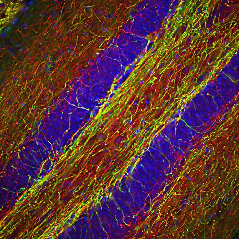
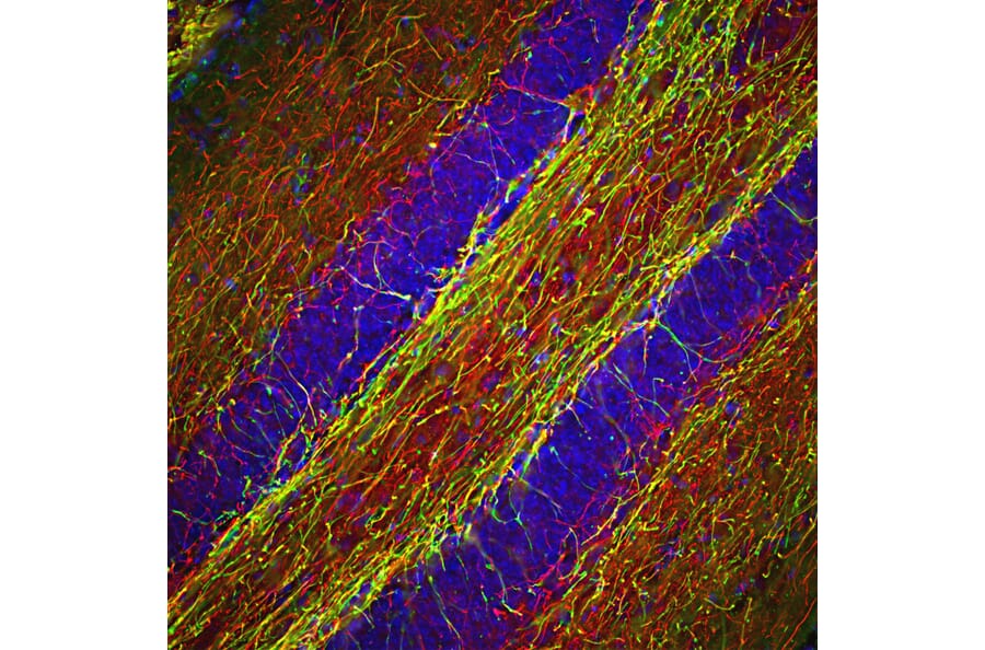
Immunohistological analysis of a mouse hippocampus section stained with Anti-NF-H Antibody, at a dilution of 1:2,000, in red, and Anti-MBP Antibody (A85322 | 1:5,000, in green. The blue is DAPI staining of nuclear DNA. Following transcardial perfusion with 4% paraformaldehyde, brain was post fixed for 24 hours, cut to 45µM, and free-floating sections were stained with the above antibodies. The Anti-NF-H Antibody labels a network of axons of different neurons, while the Anti-MBP Antibody stains myelin sheath around these axons.
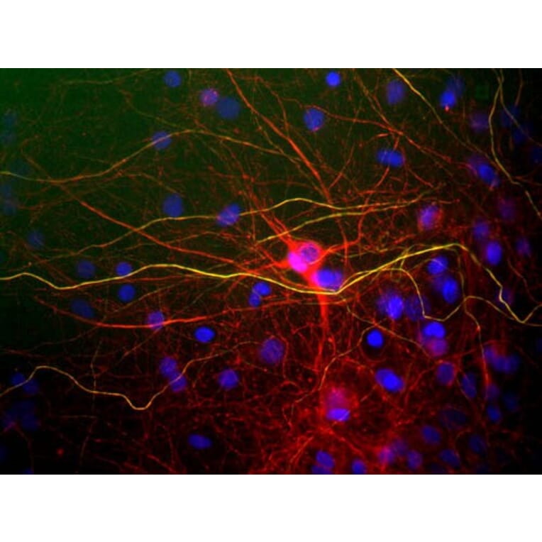
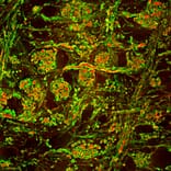
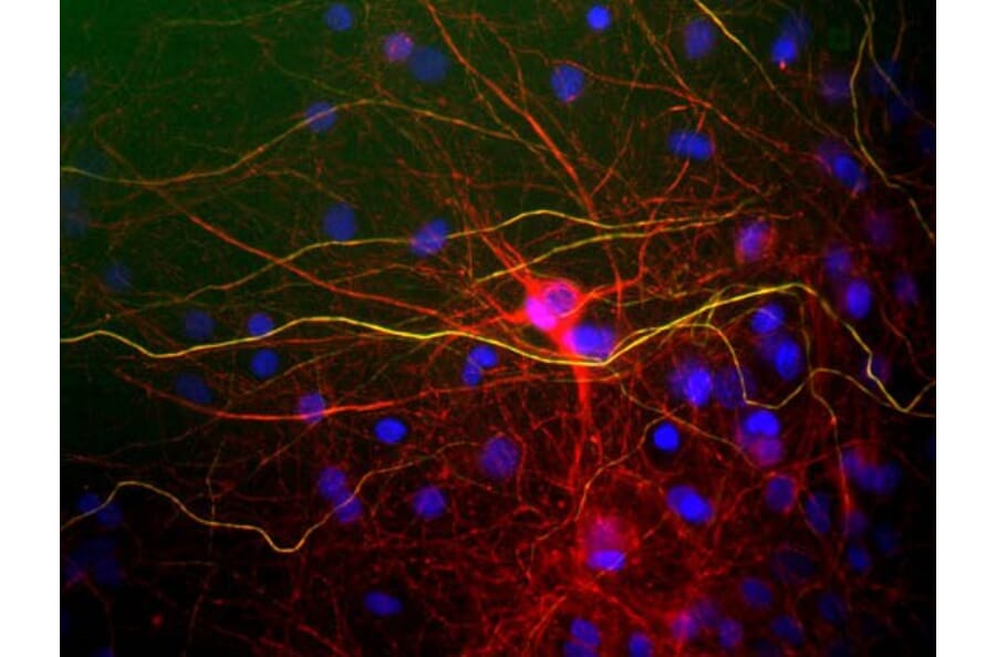
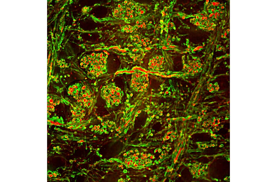
Mixed neuron/glia cultures stained with Anti-NF-H Antibody (green) and Anti-NF-L Antibody (A85286 | red). Axons contain phosphorylated NF-H and NF-L so appear yellow, while dendrites and perikarya only contain NF-L and so appear red. DNA is shown in blue.
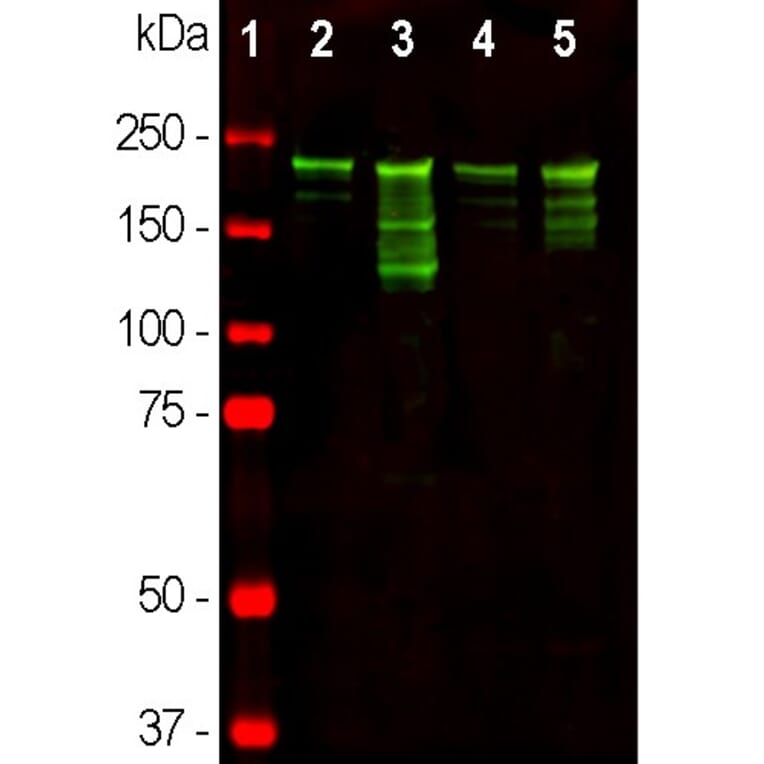
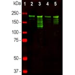
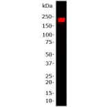
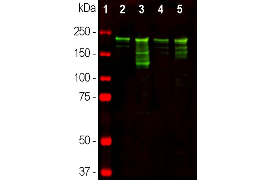
Western blot analysis of different tissue lysates using Anti-NF-H Antibody, at a dilution of 1:10,000, in green,: [Lane 1] protein standard (red), [Lane 2] rat brain, [Lane 3] rat spinal cord [Lane 4] mouse brain, and [Lane 5] mouse spinal cord lysate. Strong band at about 220kDa corresponds to the phosphorylated axonal form of the NF-H subunit. Smaller proteolytic fragments of NF-H are also detected with Anti-NF-H Antibody.
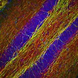
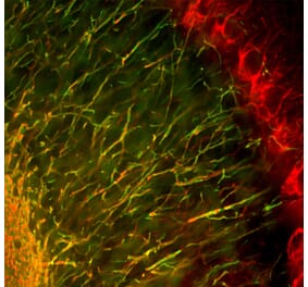
Immunofluorescent analysis of rat brain stem section stained with Anti-Myelin Basic Protein Antibody [7G7] (A85322) at a dilution of 1:5,000 (green) and costained with Anti-NF-H Antibody (A85336) at a dilution of 1:2,000 (red). This antibody stains oligodendrocytes and the myelin sheathes around axons. In this high magnification view it is clear that the NF-H antibody labels axons within the myelin sheathes.
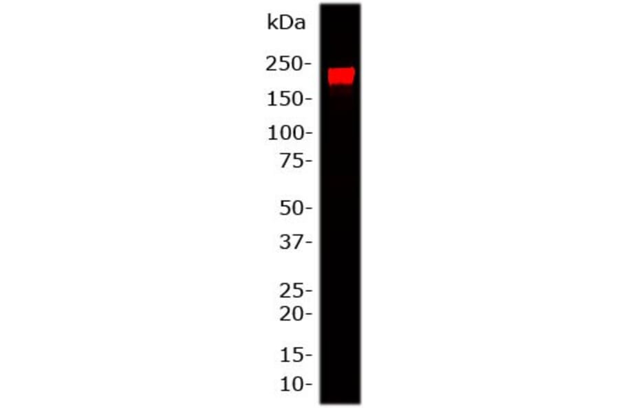
Western blot analysis of Anti-NF-H Antibody. Blot of 20 µg rat brain lysate was probed with Anti-NF-H Antibody, at a dilution of 1:25,000). A prominent band at 200 kDa corresponds to phosphorylated form of NF-H.
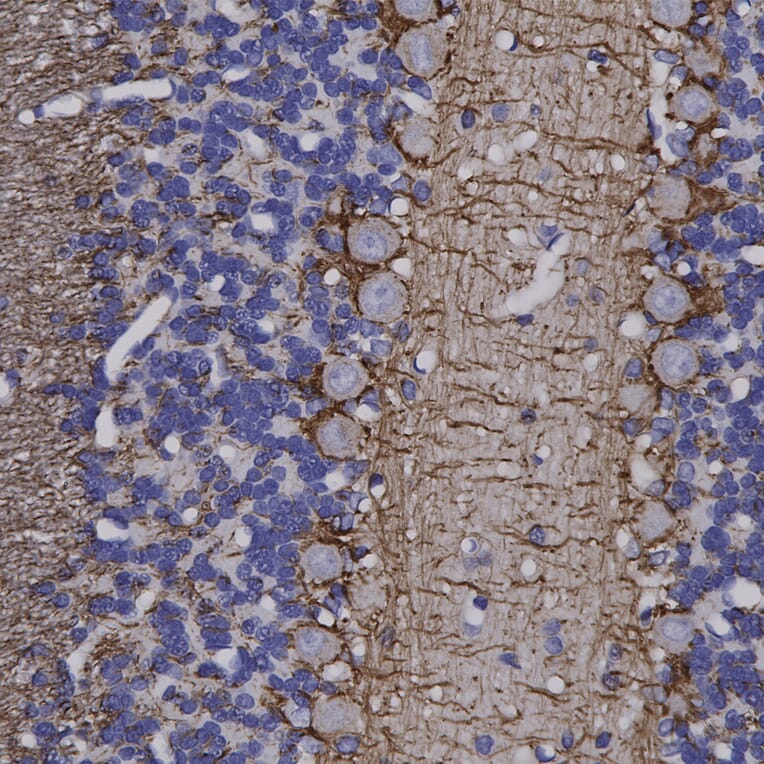
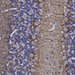
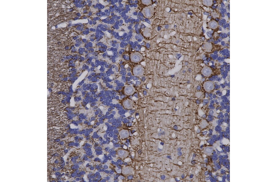
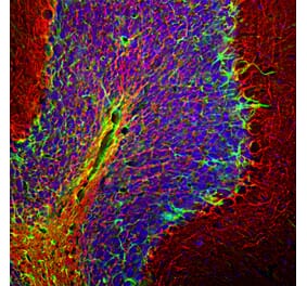
Immunohistochemistry analysis of a formalin fixed paraffin embedded rat cerebellum section with Anti-NF-H Antibody (A85336) at a dilution of 1:4,000. Anti-NF-H Antibody (A85336) labels Purkinje cell dendrites and the projections of neuronal cells within the white matter and the granular layer. Note: this antibody performs well in testing with both 4% PFA and standard NBF fixed rat, mouse and human tissues.


![Immunohistochemistry - Anti-Neurofilament Heavy Polypeptide Antibody [NF421] - BSA and Azide free (A252667) - Antibodies.com](https://cdn.antibodies.com/image/catalog/252/A252667_1.jpg?profile=product_alternative)
![Immunohistochemistry - Anti-Neurofilament Heavy Polypeptide Antibody [NE14] (A249491) - Antibodies.com](https://cdn.antibodies.com/image/catalog/249/A249491_1.jpg?profile=product_alternative)
![Immunohistochemistry - Anti-Neurofilament Heavy Polypeptide Antibody [NE14] - BSA and Azide free (A252671) - Antibodies.com](https://cdn.antibodies.com/image/catalog/252/A252671_1.jpg?profile=product_alternative)

![Immunohistochemistry - Anti-Neurofilament Heavy Polypeptide Antibody [NF421] (A249487) - Antibodies.com](https://cdn.antibodies.com/image/catalog/249/A249487_1.jpg?profile=product_alternative)
![Immunofluorescence - Anti-NF-H Antibody [9B12] (A85341) - Antibodies.com](https://cdn.antibodies.com/image/catalog/85/A85341_1.jpg?profile=product_alternative)
![Immunohistochemistry - Anti-NF-H Antibody [NAP4] (A85339) - Antibodies.com](https://cdn.antibodies.com/image/catalog/85/A85339_1.jpg?profile=product_alternative)
![Immunofluorescence - Anti-NF-H Antibody [AH1] (A85340) - Antibodies.com](https://cdn.antibodies.com/image/catalog/85/A85340_1.jpg?profile=product_alternative)
![Immunohistochemistry - Anti-Neurofilament Heavy Polypeptide Antibody [rNF421] (A249492) - Antibodies.com](https://cdn.antibodies.com/image/catalog/249/A249492_1.jpg?profile=product_alternative)