The preparation that this antibody was raised against is dominated by axonal forms of NF-H which are heavily phosphorylated on the multiple repeated NF-H KSP type sequences, and this antibody reacts very strongly with these phosphorylated repeats. Reactivity with non-phosphorylated KSP sequences is orders of magnitude weaker.
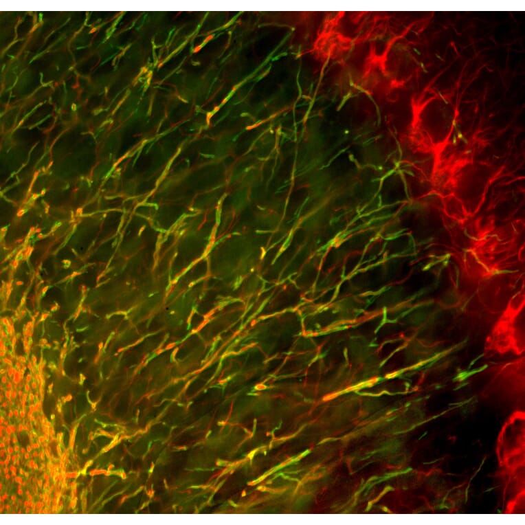
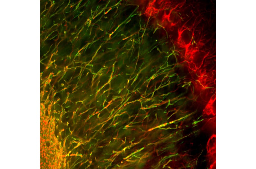
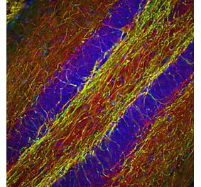
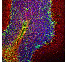
Immunofluorescent analysis of mouse cerebellum section stained with Anti-NF-H Antibody (A270594), at a dilution of 1:3,000, in red, and co-stained with Anti-Myelin Basic Protein Antibody [7G7] (A85322), at a dilution of 1:5,000, in green. Following transcardial perfusion with 4% paraformaldehyde, the mouse brain was post-fixed for 24 hours, cut to 45 µm, and free-floating sections were stained with above antibodies. Anti-NF-H Antibody (A270594) labels axons of basket and Purkinje cells and others, while Anti-Myelin Basic Protein Antibody [7G7] (A85322) stains oligodendrocyte cell bodies and the myelin sheathes around axons in the granular layer at center and the white matter at bottom left.
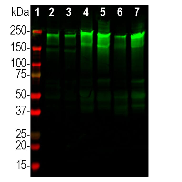
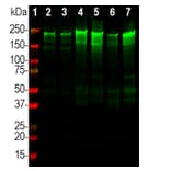
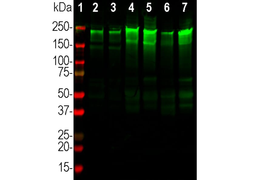
Western blot analysis of tissue lysates from different species using Anti-NF-H Antibody (A270594), at a dilution of 1:20,000, in green. The lanes contain: [Lane 1] protein standard (red), [Lane 2] rat brain, [Lane 3] mouse brain, [Lane 4] cow cerebellum, [Lane 5] cow spinal cord, [Lane 6] pig hippocampus, and [Lane 7] pig spinal cord. The strong band at about 220 kDa corresponds to the major phospho NF-H subunit. Smaller proteolytic fragments of NF-H are also detected in some preparations.
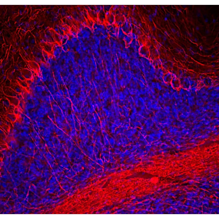
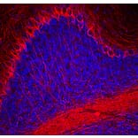
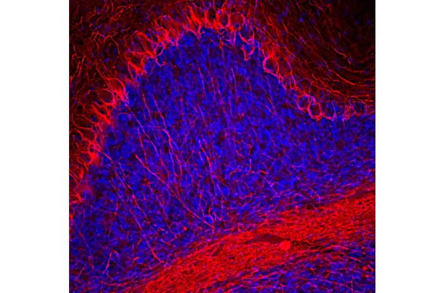
Immunofluorescence analysis of mouse cerebellum section stained with goat pAb to Anti-NF-H Antibody (A270594) at a dilution of 1:3,000 (red). Nuclei were stained with Hoechst (blue). Following transcardial perfusion with 4% paraformaldehyde, mouse brain was post fixed for 24 hours, cut to 45µM, and free-floating sections were stained with with our standard protocol. The NF-H antibody labels a network of axons of different neurons.
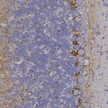
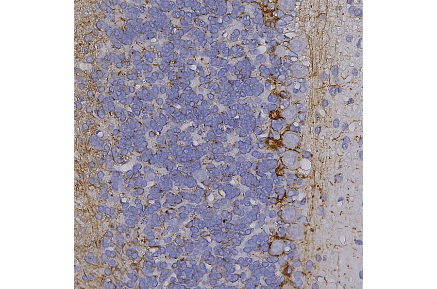
Immunohistochemistry analysis of a formalin fixed paraffin embedded rat cerebellum section with Anti-NF-H Antibody (A270594) at a dilution of 1:5,000 detected with DAB (brown) using the Vector Elite ABC-HRP detection and reagents with citra buffer retrieval. Counterstained with Hematoxylin (blue). In cerebellum, Anti-NF-H Antibody (A270594) labels networks of axonal processes and is highly expressed in basket cells.
Publishing research using Anti-NF-H Antibody (A270594)? Please let us know so that we can list the citation on this page.


![Immunohistochemistry - Anti-Neurofilament Heavy Polypeptide Antibody [NF421] - BSA and Azide free (A252667) - Antibodies.com](https://cdn.antibodies.com/image/catalog/252/A252667_1.jpg?profile=product_alternative)
![Immunohistochemistry - Anti-Neurofilament Heavy Polypeptide Antibody [NE14] (A249491) - Antibodies.com](https://cdn.antibodies.com/image/catalog/249/A249491_1.jpg?profile=product_alternative)
![Immunohistochemistry - Anti-Neurofilament Heavy Polypeptide Antibody [NE14] - BSA and Azide free (A252671) - Antibodies.com](https://cdn.antibodies.com/image/catalog/252/A252671_1.jpg?profile=product_alternative)

![Immunohistochemistry - Anti-Neurofilament Heavy Polypeptide Antibody [NF421] (A249487) - Antibodies.com](https://cdn.antibodies.com/image/catalog/249/A249487_1.jpg?profile=product_alternative)
![Immunofluorescence - Anti-NF-H Antibody [9B12] (A85341) - Antibodies.com](https://cdn.antibodies.com/image/catalog/85/A85341_1.jpg?profile=product_alternative)
![Immunohistochemistry - Anti-NF-H Antibody [NAP4] (A85339) - Antibodies.com](https://cdn.antibodies.com/image/catalog/85/A85339_1.jpg?profile=product_alternative)
![Immunofluorescence - Anti-NF-H Antibody [AH1] (A85340) - Antibodies.com](https://cdn.antibodies.com/image/catalog/85/A85340_1.jpg?profile=product_alternative)
![Immunohistochemistry - Anti-Neurofilament Heavy Polypeptide Antibody [NF421 + NFL/736] (A250886) - Antibodies.com](https://cdn.antibodies.com/image/catalog/250/A250886_1.jpg?profile=product_alternative)
![Immunohistochemistry - Anti-Neurofilament Heavy Polypeptide Antibody [NEFL.H/2324R] - BSA and Azide free (A252673) - Antibodies.com](https://cdn.antibodies.com/image/catalog/252/A252673_1.jpg?profile=product_alternative)