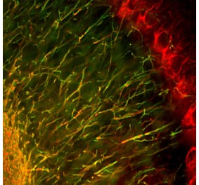
Culture from E20 rat cortex grown in culture and stained with Anti-NF-H Antibody [9B12] (A85341) at a dilution of 1:5,000 (red) and also with Anti-NF-L Antibody (A85451) at a dilution of 1:5,000 (green). The Anti-NF-H Antibody [9B12] (A85341) antibody bind to phosphorylated NF-H found in some mature axonal profiles, while the NF-L binds to both axonal, dendritic and developing neuronal processes. The mature axonal profiles contain both NF-L and phosphorylated NF-H and so appear yellow, while processes expressing only NF-L appear green. Nuclei were stained with DAPI (blue).
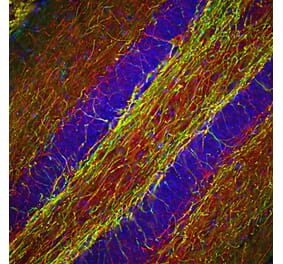
Mixed neuron/glial cultures stained with Anti-NF-H Antibody (red) and Anti-GFAP Antibody (A85419 | green). Axonal profiles are stained in red, while astrocytic cells are revealed in green. Nuclei are revealed with a fluorescent DNA stain (blue).
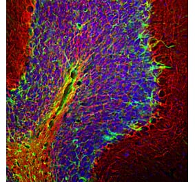
Immunofluorescent analysis of a rat brain coronal section of the third ventricle stained with Anti-NF-H Antibody [9B12] (A85341), at a dilution of 1:5,000, in green. The nuclear DNA is visualised in blue using Hoechst staining. Following transcardial perfusion with 4% paraformaldehyde, the brain was post-fixed for 24 hours, cut to 45 µm, and free-floating sections were stained with the above antibody. The Anti-NF-H Antibody [9B12] (A85341) is a robust marker of the axons of neuronal cells.
Western blot analysis of different tissue lysates using Anti-NF-H Antibody [9B12] (A85341), at a dilution of 1:10,000, in green. The lanes contain samples of: [Lane 1] Protein standards, [Lane 2] rat spinal cord [Lane 3] mouse spinal cord, and [Lane 4] cow spinal cord. The strong band at about 200-220 kDa corresponds to the major phosphorylated form of the NF-H subunit. Smaller proteolytic fragments of NF-H are also detected in some preparations.
Strip blots of crude rat spinal cord extract stained with three different antibodies to phosphorylated NF-H: Lane 1: Anti-NF-H Antibody (A85339). Lane 2: Anti-NF-H Antibody (A85338). Lane 3: Anti-NF-H Antibody (A85341). All three antibodies bind to a prominent band with an apparent SDS-PAGE molecular weight of 200 kDa.
Immunohistochemistry analysis of a formalin fixed paraffin embedded human cerebellum section with Anti-NF-H Antibody [9B12] (A85341) at a dilution of 1:10,000. Anti-NF-H Antibody [9B12] (A85341) labels Purkinje cell dendrites and the projections of neuronal cells within the granular layer. Note: this antibody performs well in testing with both 4% PFA and standard NBF fixed rat, mouse and human tissues.


![Immunofluorescence - Anti-NF-H Antibody [9B12] (A85341) - Antibodies.com](https://cdn.antibodies.com/image/catalog/85/A85341_1.jpg?profile=product_top)
![Immunofluorescence - Anti-NF-H Antibody [9B12] (A85341) - Antibodies.com](https://cdn.antibodies.com/image/catalog/85/A85341_2.jpg?profile=product_top)
![Immunofluorescence - Anti-NF-H Antibody [9B12] (A85341) - Antibodies.com](https://cdn.antibodies.com/image/catalog/85/A85341_3.jpg?profile=product_top)
![Western Blot - Anti-NF-H Antibody [9B12] (A85341) - Antibodies.com](https://cdn.antibodies.com/image/catalog/85/A85341_4.jpg?profile=product_top)
![Western Blot - Anti-NF-H Antibody [9B12] (A85341) - Antibodies.com](https://cdn.antibodies.com/image/catalog/85/A85341_5.jpg?profile=product_top)
![Immunohistochemistry - Anti-NF-H Antibody [9B12] (A85341) - Antibodies.com](https://cdn.antibodies.com/image/catalog/85/A85341_6.jpg?profile=product_top)
![Immunofluorescence - Anti-NF-H Antibody [9B12] (A85341) - Antibodies.com](https://cdn.antibodies.com/image/catalog/85/A85341_1.jpg?profile=product_top_thumb)
![Immunofluorescence - Anti-NF-H Antibody [9B12] (A85341) - Antibodies.com](https://cdn.antibodies.com/image/catalog/85/A85341_2.jpg?profile=product_top_thumb)
![Immunofluorescence - Anti-NF-H Antibody [9B12] (A85341) - Antibodies.com](https://cdn.antibodies.com/image/catalog/85/A85341_3.jpg?profile=product_top_thumb)
![Western Blot - Anti-NF-H Antibody [9B12] (A85341) - Antibodies.com](https://cdn.antibodies.com/image/catalog/85/A85341_4.jpg?profile=product_top_thumb)
![Western Blot - Anti-NF-H Antibody [9B12] (A85341) - Antibodies.com](https://cdn.antibodies.com/image/catalog/85/A85341_5.jpg?profile=product_top_thumb)
![Immunohistochemistry - Anti-NF-H Antibody [9B12] (A85341) - Antibodies.com](https://cdn.antibodies.com/image/catalog/85/A85341_6.jpg?profile=product_top_thumb)
![Immunofluorescence - Anti-NF-H Antibody [9B12] (A85341) - Antibodies.com](https://cdn.antibodies.com/image/catalog/85/A85341_1.jpg?profile=product_image)
![Immunofluorescence - Anti-NF-H Antibody [9B12] (A85341) - Antibodies.com](https://cdn.antibodies.com/image/catalog/85/A85341_2.jpg?profile=product_image)
![Immunofluorescence - Anti-NF-H Antibody [9B12] (A85341) - Antibodies.com](https://cdn.antibodies.com/image/catalog/85/A85341_3.jpg?profile=product_image)
![Western Blot - Anti-NF-H Antibody [9B12] (A85341) - Antibodies.com](https://cdn.antibodies.com/image/catalog/85/A85341_4.jpg?profile=product_image)
![Western Blot - Anti-NF-H Antibody [9B12] (A85341) - Antibodies.com](https://cdn.antibodies.com/image/catalog/85/A85341_5.jpg?profile=product_image)
![Immunohistochemistry - Anti-NF-H Antibody [9B12] (A85341) - Antibodies.com](https://cdn.antibodies.com/image/catalog/85/A85341_6.jpg?profile=product_image)
![Immunohistochemistry - Anti-Neurofilament Heavy Polypeptide Antibody [NF421] - BSA and Azide free (A252667) - Antibodies.com](https://cdn.antibodies.com/image/catalog/252/A252667_1.jpg?profile=product_alternative)
![Immunohistochemistry - Anti-Neurofilament Heavy Polypeptide Antibody [NE14] - BSA and Azide free (A252671) - Antibodies.com](https://cdn.antibodies.com/image/catalog/252/A252671_1.jpg?profile=product_alternative)
![Immunohistochemistry - Anti-Neurofilament Heavy Polypeptide Antibody [NE14] (A249491) - Antibodies.com](https://cdn.antibodies.com/image/catalog/249/A249491_1.jpg?profile=product_alternative)

![Immunohistochemistry - Anti-Neurofilament Heavy Polypeptide Antibody [NF421] (A249487) - Antibodies.com](https://cdn.antibodies.com/image/catalog/249/A249487_1.jpg?profile=product_alternative)
![Immunohistochemistry - Anti-NF-H Antibody [NAP4] (A85339) - Antibodies.com](https://cdn.antibodies.com/image/catalog/85/A85339_1.jpg?profile=product_alternative)
![Immunofluorescence - Anti-NF-H Antibody [AH1] (A85340) - Antibodies.com](https://cdn.antibodies.com/image/catalog/85/A85340_1.jpg?profile=product_alternative)
![Immunohistochemistry - Anti-Neurofilament Heavy Polypeptide Antibody [rNF421] (A249492) - Antibodies.com](https://cdn.antibodies.com/image/catalog/249/A249492_1.jpg?profile=product_alternative)
![Immunohistochemistry - Anti-Neurofilament Heavy Polypeptide Antibody [rNF421] - BSA and Azide free (A252672) - Antibodies.com](https://cdn.antibodies.com/image/catalog/252/A252672_1.jpg?profile=product_alternative)