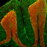
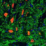
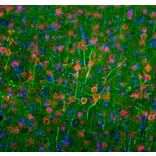
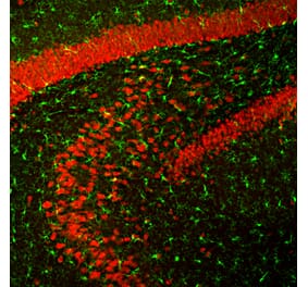
Immunofluorescent analysis of a section of adult mouse cerebellum stained with Anti-NeuN Antibody, at a dilution of 1:5,000, in red, and Anti-GFAP Antibody (A85307 | 1:5,000, in green. Following transcardial perfusion of mouse with 4% paraformaldehyde, brain was post fixed for 24 hours, cut to 45µM, and free-floating sections were stained with the above antibodies. The Anti-NeuN Antibody stains the nuclei of neurons in the cerebellar granule layer. The Anti-GFAP Antibody stains the processes of Bergmann glia in the molecular layer and astroglia in the granule and white matter layers.
Paraformaldehyde fixed frozen section of adult rat brain stem stained with Anti-NeuN Antibody (red) and Anti-MAP2 Antibody (A85296 | green). DNA is shown in blue. Note, that the Anti-NeuN Antibody stains neuronal nuclei and distal perikarya and that the Anti-MAP2 Antibody stains the dendrites extending from these cells. Anti-NeuN Antibody stains exactly like the original NeuN monoclonal antibody. These antibodies do not bind to the nuclei of perikarya of any non-neuronal cells, so that they can be used to identify and quantify neurons.
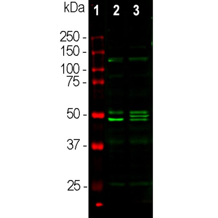
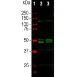
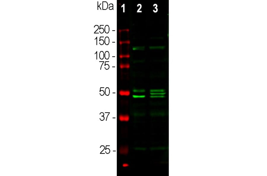
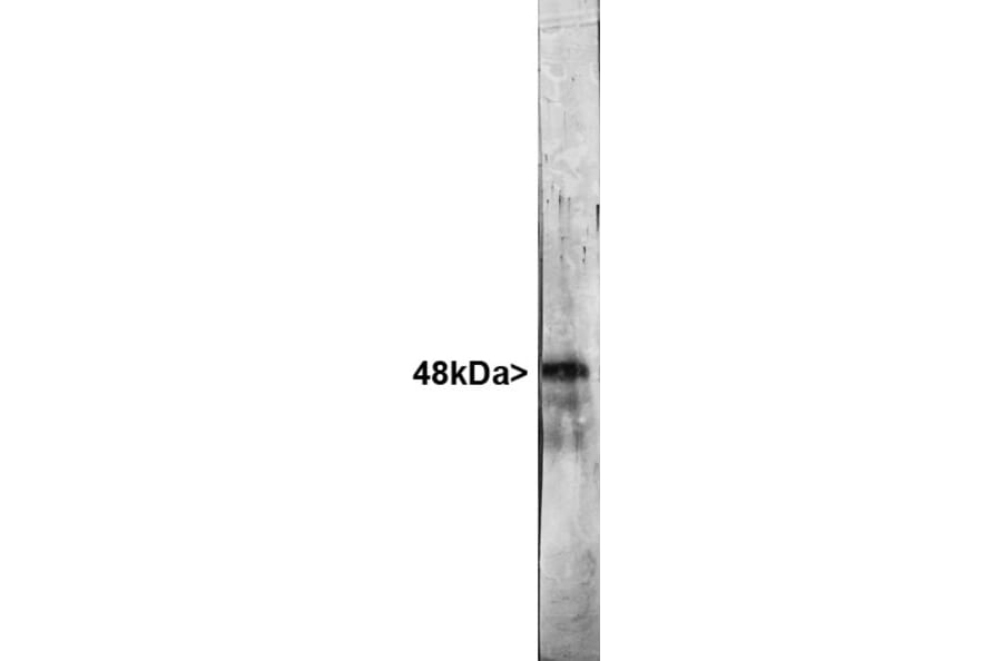
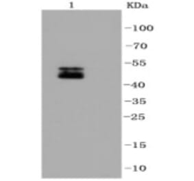
Western blot analysis of whole brain lysates using Anti-NeuN Antibody, at a dilution of 1:1,000, in green,: [Lane 1] protein standard (red), [Lane 2] mouse brain, [Lane 3] rat brain. Bands at about 48 kDa correspond to protein isotypes of NeuN.
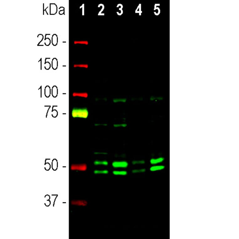
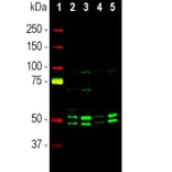
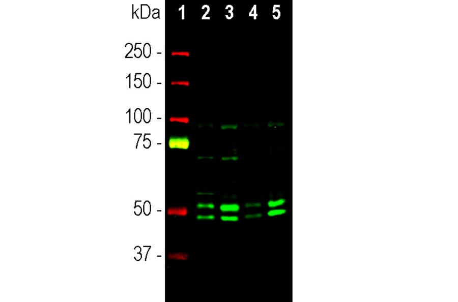
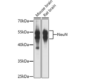
Western blot analysis of cytosolic (cyt) and nuclear enriched (nuc) fractions of whole brain lysates using Anti-NeuN Antibody (A85403), at a dilution of 1:1,000, in green. The lanes contain samples of: [Lane 1] Protein standards, in red, [Lane 2] rat cyt, [Lane 3] rat nuc, [Lane 4] mouse cyt, and [Lane 5] mouse nuc lysate. Two bands of 46 and 48kDa correspond to the two alternate transcripts of the NeuN protein. Western blot was performed under non-reducing conditions.
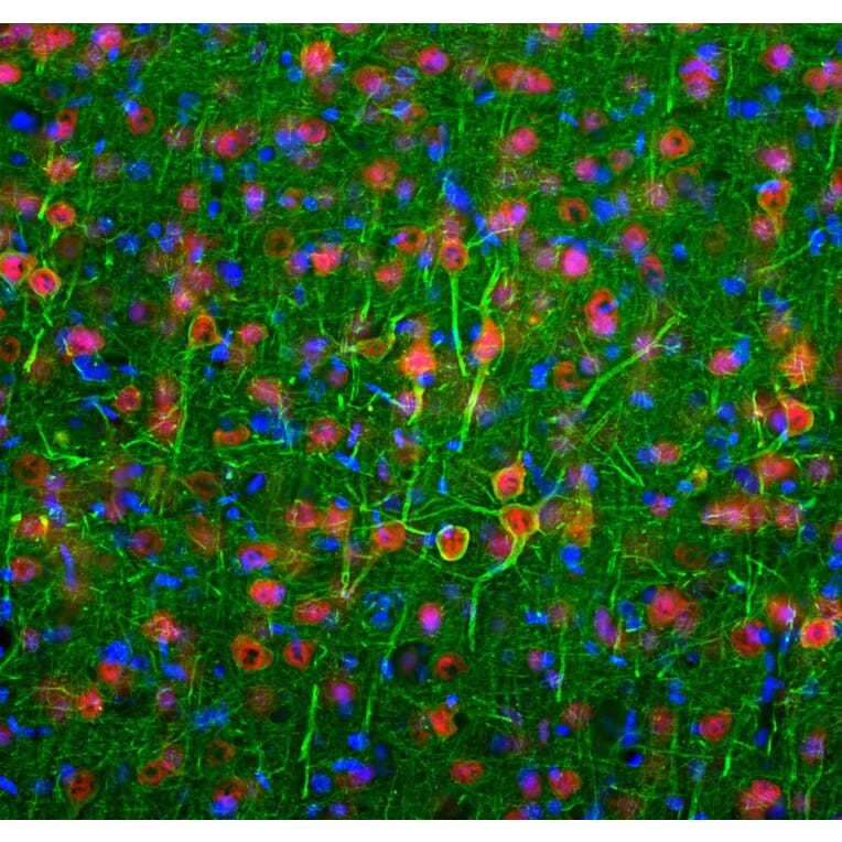
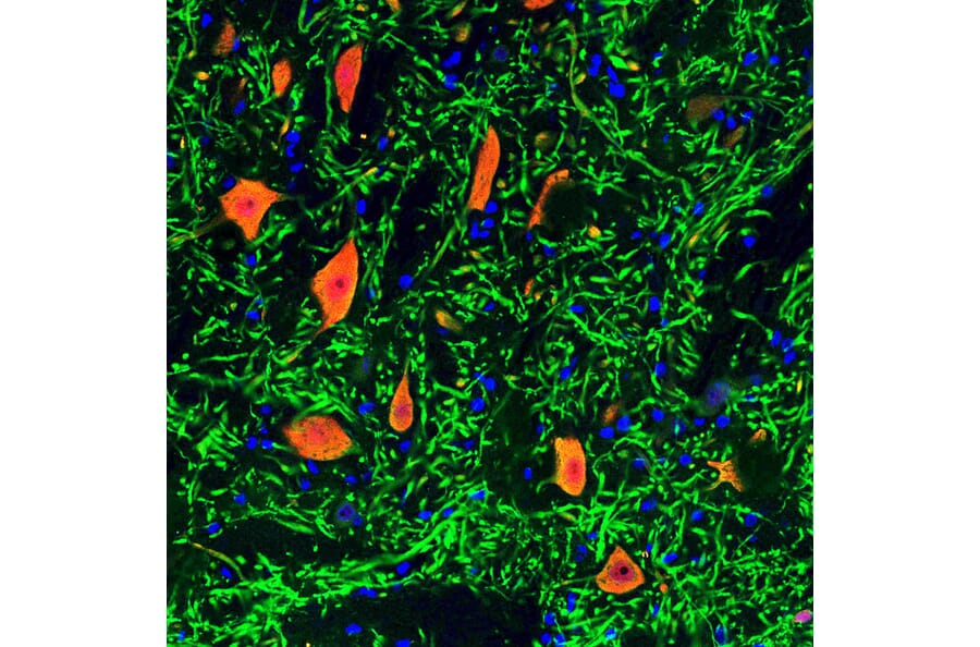
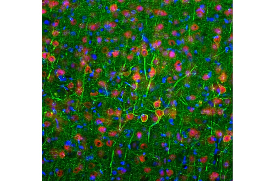
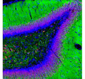
Immunofluorescent analysis of a rat brain section stained with Anti-NeuN Antibody (A85403), at a dilution of 1:2,000, in red, and co-stained with Anti-MAP2 Antibody (A85363), at a dilution of 1:5,000, in green. Following transcardial perfusion of a rat with 4% paraformaldehyde, the brain was post-fixed for 24 hours, cut to 45 µm, and free-floating sections were stained with the above antibodies. The Anti-NeuN Antibody (A85403) selectively stains nuclei and cytoplasm of neuronal cells, while the Anti-MAP2 Antibody (A85363) labels dendrites and overlaps with Fox3 staining in neuronal perikarya.
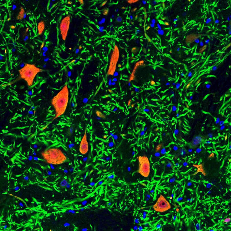
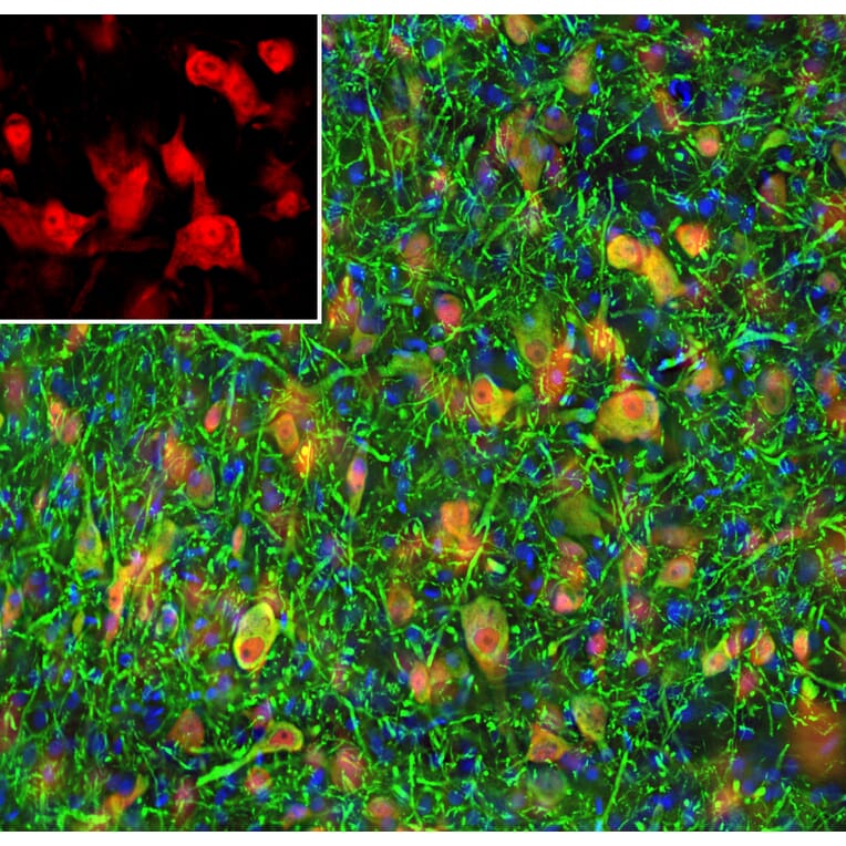
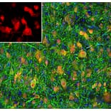
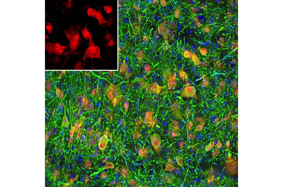
Immunofluorescent analysis of a rat frontal cortex section stained with Anti-NF-L Antibody [6H112] (A270560) at a dilution of 1:2,000 (green) and costained with Anti-NeuN Antibody (A85403) at a dilution of 1:2,000 (red). Following transcardial perfusion of rat with 4% paraformaldehyde, brain was post fixed for 24 hours, cut to 45µM, and free-floating sections were stained with the above antibodies. The Anti-NF-L Antibody [6H112] (A270560) labels the cell bodies and processes of pyramidal neurons, as well as dendrites and axons of other neuronal cells, while the FOX3/NeuN antibody selectively stains nuclei and cytoplasm of neuronal cells.
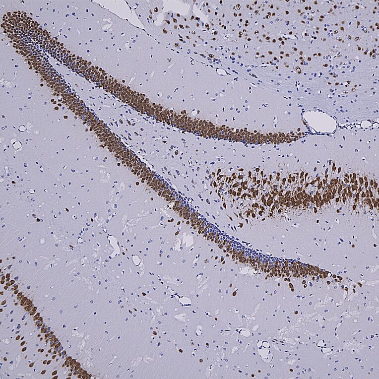
Immunohistochemistry analysis of a formalin fixed paraffin embedded mouse hippocampus section with Anti-NeuN Antibody (A85403) at a dilution of 1:4,000 detected with DAB (brown) using the Vector Labs ImmPRESS method and reagents with citra buffer retrieval. The Anti-NeuN Antibody (A85403) selectively labels the nuclei and distal perikaryal of most neuronal cell populations. Note: this antibody performs well in testing with 4% PFA and NBF fixed mouse, human, and rat tissues.


![Immunofluorescence - Anti-NeuN Antibody [1B7] (A85405) - Antibodies.com](https://cdn.antibodies.com/image/catalog/85/A85405_1.jpg?profile=product_alternative)
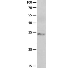
![Western Blot - Anti-NeuN Antibody [ARC0202] (A306978) - Antibodies.com](https://cdn.antibodies.com/image/catalog/306/A306978_1.jpg?profile=product_alternative)
![Immunohistochemistry - Anti-NeuN Antibody [NeuN/288R] (A277965) - Antibodies.com](https://cdn.antibodies.com/image/catalog/277/A277965_1.jpg?profile=product_alternative)
![Immunohistochemistry - Anti-NeuN Antibody [NeuN/288R] - BSA and Azide free (A278553) - Antibodies.com](https://cdn.antibodies.com/image/catalog/278/A278553_1.jpg?profile=product_alternative)
![Immunohistochemistry - Anti-NeuN Antibody [NeuN/6694R] - BSA and Azide free (A278554) - Antibodies.com](https://cdn.antibodies.com/image/catalog/278/A278554_1.jpg?profile=product_alternative)
![Immunohistochemistry - Anti-NeuN Antibody [NeuN/6694R] (A277966) - Antibodies.com](https://cdn.antibodies.com/image/catalog/277/A277966_1.jpg?profile=product_alternative)
![Immunohistochemistry - Anti-NeuN Antibody [NeuN/7071R] (A277967) - Antibodies.com](https://cdn.antibodies.com/image/catalog/277/A277967_1.jpg?profile=product_alternative)
![Immunohistochemistry - Anti-NeuN Antibody [RM312] (A121402) - Antibodies.com](https://cdn.antibodies.com/image/catalog/121/A121385_1.png?profile=product_alternative)