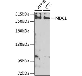
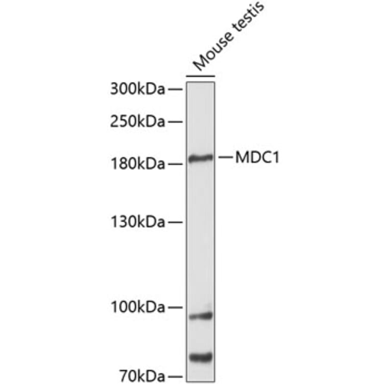
Western blot analysis of extracts of mouse testis, using Anti-MDC1 Antibody (A88650) at 1:3,000 dilution. The secondary antibody was Goat Anti-Rabbit IgG H&L Antibody (HRP) at 1:10,000 dilution. Lysates/proteins were present at 25µg per lane. The blocking buffer used was 3% non-fat dry milk in TBST. Detection was with a ECL Basic Kit. Exposure time: 90s.
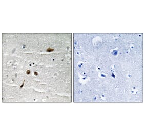
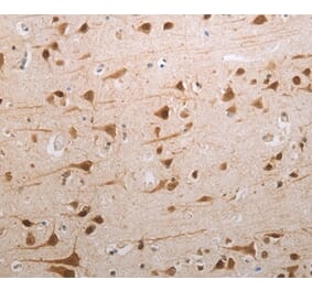
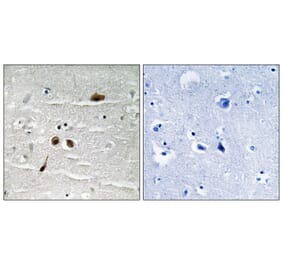
Immunohistochemistry analysis of paraffin-embedded human esophageal cancer using Anti-MDC1 Antibody (A88650) at a dilution of 1:100 (40x lens). Perform high pressure antigen retrieval with 10 mM citrate buffer pH 6.0 before commencing with IHC staining protocol.
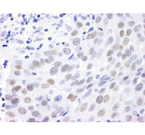
Immunohistochemistry analysis of paraffin-embedded mouse testis using Anti-MDC1 Antibody (A88650) at a dilution of 1:100 (40x lens). Perform high pressure antigen retrieval with 10 mM citrate buffer pH 6.0 before commencing with IHC staining protocol.
Immunofluorescence analysis of U-2 OS cells using Anti-MDC1 Antibody (A88650) at a dilution of 1:100 (40x lens). DAPI was used to stain the cell nuclei (blue).
Publishing research using Anti-MDC1 Antibody (A88650)? Please let us know so that we can list the citation on this page.
Alternative products to Anti-MDC1 Antibody (A88650)


![Western Blot - Anti-MDC1 Antibody [P2B11] (A304945) - Antibodies.com](https://cdn.antibodies.com/image/catalog/304/A304945_1.png?profile=product_alternative)