The epitope of this antibody has been mapped to the peptide IKQRLKLKDGGHYDAEVKTT, with the central KLKDGGHYDA sequence a key component of antibody binding.
Applications
WB, ICC/IF, IHC
Dilutions
WB: 1:2,000, ICC/IF: 1:500, IHC: 1:2,000
Immunogen
Recombinant full-length mCherry, expressed in and purified from E. coli.
Supplied in Phosphate Buffered Saline with 50% Glycerol and 5mM Sodium Azide.
Storage
Shipped at 4°C. Upon delivery aliquot and store at -20°C. Avoid freeze/thaw cycles.
General Notes
This antibody can be used to verify the size of fusion constructs by western blotting, and to amplify the endogenous fluorescence of mCherry in transfected cells.
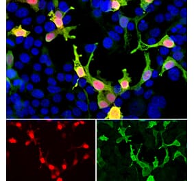
Immunofluorescent analysis of HEK293 cells stably transduced with a lentiviral vector expressing an mCherry-HA construct (red) and stained with Anti-mCherry Antibody [5A6] (A104343), at a dilution of 1:500, in green. The blue is Hoechst staining of nuclear DNA. Anti-mCherry Antibody [5A6] (A104343) reveals the mCherry protein expressed only in transduced cells which appear golden in color. Untransduced cells expressing no mCherry are not recognized by Anti-mCherry Antibody [5A6] (A104343) and so only their nuclei are visible.
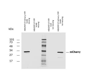
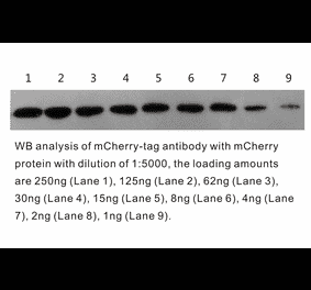
Western Blot - Anti-mCherry Antibody [5A6] (A104343)
Western blot analysis of various HEK293 cell lysates using Anti-mCherry Antibody [5A6] (A104343), at a dilution of 1:5,000, in green. The lanes contain: [Lane 1] protein standard, [Lane 2] untransfected HEK293 lysate, [Lane 3] lysate of HEK293 cells transfected with mCherry-HA construct, and [Lane 4] lysate of HEK293 cells transfected with an acGFP construct. As expected from our epitope mapping data, Anti-mCherry Antibody [5A6] (A104343) does not recognize eGFP. The major band at about 28kDa corresponds to the full length mCherry protein and the lower band at about 21kDa is an mCherry breakdown product.
Immunohistochemistry analysis of formalin fixed paraffin embedded mCherry transfected monkey brain with Anti-mCherry Antibody [5A6] (A104343) at a dilution of 1:2,000 detected with DAB (brown) following the Vector labs Vector Labs ImmPRESS method and reagents with citra buffer retrieval. Counterstained with Hematoxylin (blue). Anti-mCherry Antibody [5A6] (A104343) specifically detected the soma and axons of mCherry positive neurons in the cerebellum.
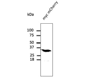
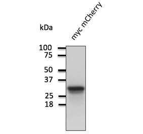
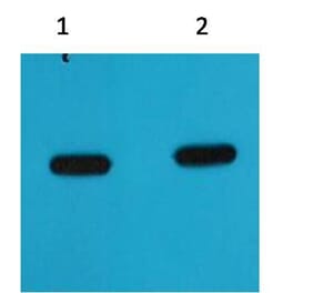
Western Blot - Anti-mCherry Antibody [5A6] (A104343)
Western blot analysis of HEK293 whole cell lysates using Anti-mCherry Antibody [5A6] (A104343) at a dilution of 1:2,000, (green). The lanes contain: [Lane 1] protein standard, [Lane 2] HEK293, [Lane 3] HEK293 cells stably transduced with lenti-virus containing mCherry-HA construct, and [Lane 4] HEK293 cells transfected with mCherry-HAI construct. Major band at about 28kDa corresponds to mCherry protein. The band at 20kDa corresponds to cleaved form of mCherry protein.
Publishing research using Anti-mCherry Antibody [5A6] (A104343)? Please let us know so that we can list the citation on this page.


![Immunofluorescence - Anti-mCherry Antibody [5A6] (A104343) - Antibodies.com](https://cdn.antibodies.com/image/catalog/104/A104343_1.jpg?profile=product_top)
![Western Blot - Anti-mCherry Antibody [5A6] (A104343) - Antibodies.com](https://cdn.antibodies.com/image/catalog/104/A104343_2.jpg?profile=product_top)
![Immunohistochemistry - Anti-mCherry Antibody [5A6] (A104343) - Antibodies.com](https://cdn.antibodies.com/image/catalog/104/A104343_3.jpg?profile=product_top)
![Western Blot - Anti-mCherry Antibody [5A6] (A104343) - Antibodies.com](https://cdn.antibodies.com/image/catalog/104/A104343_4.jpg?profile=product_top)
![Immunofluorescence - Anti-mCherry Antibody [5A6] (A104343) - Antibodies.com](https://cdn.antibodies.com/image/catalog/104/A104343_1.jpg?profile=product_top_thumb)
![Western Blot - Anti-mCherry Antibody [5A6] (A104343) - Antibodies.com](https://cdn.antibodies.com/image/catalog/104/A104343_2.jpg?profile=product_top_thumb)
![Immunohistochemistry - Anti-mCherry Antibody [5A6] (A104343) - Antibodies.com](https://cdn.antibodies.com/image/catalog/104/A104343_3.jpg?profile=product_top_thumb)
![Western Blot - Anti-mCherry Antibody [5A6] (A104343) - Antibodies.com](https://cdn.antibodies.com/image/catalog/104/A104343_4.jpg?profile=product_top_thumb)
![Immunofluorescence - Anti-mCherry Antibody [5A6] (A104343) - Antibodies.com](https://cdn.antibodies.com/image/catalog/104/A104343_1.jpg?profile=product_image)
![Western Blot - Anti-mCherry Antibody [5A6] (A104343) - Antibodies.com](https://cdn.antibodies.com/image/catalog/104/A104343_2.jpg?profile=product_image)
![Immunohistochemistry - Anti-mCherry Antibody [5A6] (A104343) - Antibodies.com](https://cdn.antibodies.com/image/catalog/104/A104343_3.jpg?profile=product_image)
![Western Blot - Anti-mCherry Antibody [5A6] (A104343) - Antibodies.com](https://cdn.antibodies.com/image/catalog/104/A104343_4.jpg?profile=product_image)

![Immunohistochemistry - Anti-mCherry Antibody [1C51] (A85305) - Antibodies.com](https://cdn.antibodies.com/image/catalog/85/A85305_1.jpg?profile=product_alternative)

![Dose-response - Anti-mCherry Nanobody [SAA0406] (A337467) - Antibodies.com](https://cdn.antibodies.com/image/catalog/337/A337467_1.jpg?profile=product_alternative)
![SDS-PAGE - Anti-mCherry Nanobody [SAA1128] (A337479) - Antibodies.com](https://cdn.antibodies.com/image/catalog/337/A337479_1.jpg?profile=product_alternative)

![SDS-PAGE - Anti-mCherry Nanobody [SAA0879] (A337478) - Antibodies.com](https://cdn.antibodies.com/image/catalog/337/A337478_1.jpg?profile=product_alternative)