Supplied in Phosphate Buffered Saline with 50% Glycerol and 5mM Sodium Azide.
Storage
Shipped at 4°C. Upon delivery aliquot and store at -20°C. Avoid freeze/thaw cycles.
General Notes
This antibody can be used to verify the size of fusion constructs by western blotting, and to amplify the endogenous fluorescence of mCherry in transfected cells.
Immunohistochemistry analysis of formalin fixed paraffin embedded mCherry transfected monkey brain with Anti-mCherry Antibody [1C51] (A85305) at a dilution of 1:2,000 detected in DAB (brown) following the ImmPress method. Counterstained with Hematoxylin (blue). Anti-mCherry Antibody [1C51] (A85305) specifically detected the soma and axons of mCherry positive neurons in the cortex.
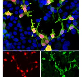
HEK293 cells transfected with mCherry and visualized in red. The cells were stained with Anti-mCherry in the green channel, and visualized using a confocal microscope. Transfected cells are yellow, showing overlap of the mCherry and the Anti-mCherry Antibody. Untransfected HEK293 cells do not express Cherry and do not stain with the antibody, but their nuclei can be visualized using a DNA stain (blue).
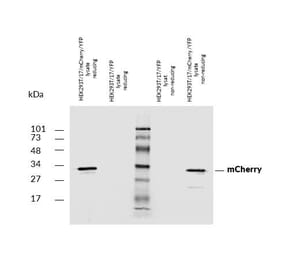
Western Blot - Anti-mCherry Antibody [1C51] (A85305)
Western blot analysis of HEK293 cell lysates, and recombinant protein solutions using Anti-mCherry Antibody [1C51] (A85305), at a dilution of 1:1,000, in green. The lanes contain samples of: [Lane 1] Protein standards, [Lane 2] HEK293 cells, [Lane 3] HEK293 cells transfected with an mCherry-HA construct, [Lane 4] mCherry recombinant protein, [Lane 5] GFP recombinant protein, and [Lane 6] HEK293 cells transfected with a GFP construct. The major band at about 30 kDa corresponds to mCherry protein. The Anti-mCherry Antibody [1C51] (A85305) does not react with GFP protein. The same blot was simultaneously probed with Anti-Heat Shock Protein 60 Antibody (A85438), at a dilution of 1:5,000, in red, which shows a band at 60 kDa seen only in cell lysates as a positive loading control.
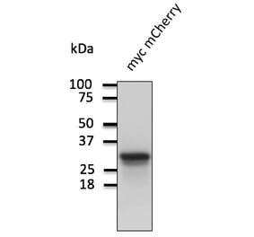
Western Blot - Anti-mCherry Antibody [1C51] (A85305)
Western blot analysis of HEK293 cell lysates using Anti-mCherry Antibody [1C51] (A85305) at a dilution of 1:2,000, (green), and rabbit pAb to GAPDH, RPCA-GAPDH at a dilution of 1:5,000 (red). The lanes contain: [Lane 1] protein molecular weight standard of indicated molecular weight in kDa, [Lane 2] non transfected HEK293 control cells, [Lane 3] HEK293 cells transfected with pCI-Neo-mod vector expressing tdTomato protein, [Lane 4] HEK293 cells transfected with pCI-Neo-mod vector expressing mCherry-HA protein. †he Anti-mCherry Antibody [1C51] (A85305) antibody recognizes tdTomato and mCherry proteins revealing major bands at about 60kDa and 30kDa, (green), respectfully. The tdTomato construct tested is identical to that found in several widely used expression vectors and contains two identical fluorescent modules, explaining why it is twice a large as the mCherry construct which contains only one fluorescent module. The red band at 37kDa corresponds to GAPDH protein used as a loading control.
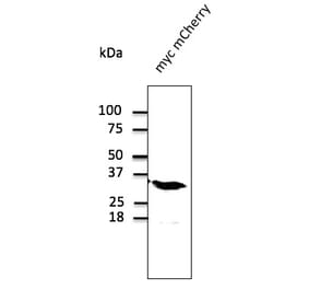
Western Blot - Anti-mCherry Antibody [1C51] (A85305)
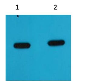
Blot of crude extract of HEK293 cells transfected with pFin-EF1-mCherry vector in lane labelled “+”. The “-” lane is a blot of an equal amount of protein extract from untransfected HEK293 cells. Anti-mCherry Antibody binds a major band running at ~28 kDa corresponding to intact full-length mCherry. The two other bands are clearly processed forms of mCherry as they are not present in non-transfected HEK293 cells.


![Immunohistochemistry - Anti-mCherry Antibody [1C51] (A85305) - Antibodies.com](https://cdn.antibodies.com/image/catalog/85/A85305_1.jpg?profile=product_top)
![Immunofluorescence - Anti-mCherry Antibody [1C51] (A85305) - Antibodies.com](https://cdn.antibodies.com/image/catalog/85/A85305_2.jpg?profile=product_top)
![Western Blot - Anti-mCherry Antibody [1C51] (A85305) - Antibodies.com](https://cdn.antibodies.com/image/catalog/85/A85305_3.jpg?profile=product_top)
![Western Blot - Anti-mCherry Antibody [1C51] (A85305) - Antibodies.com](https://cdn.antibodies.com/image/catalog/85/A85305_4.jpg?profile=product_top)
![Western Blot - Anti-mCherry Antibody [1C51] (A85305) - Antibodies.com](https://cdn.antibodies.com/image/catalog/85/A85305_5.jpg?profile=product_top)
![Immunohistochemistry - Anti-mCherry Antibody [1C51] (A85305) - Antibodies.com](https://cdn.antibodies.com/image/catalog/85/A85305_1.jpg?profile=product_top_thumb)
![Immunofluorescence - Anti-mCherry Antibody [1C51] (A85305) - Antibodies.com](https://cdn.antibodies.com/image/catalog/85/A85305_2.jpg?profile=product_top_thumb)
![Western Blot - Anti-mCherry Antibody [1C51] (A85305) - Antibodies.com](https://cdn.antibodies.com/image/catalog/85/A85305_3.jpg?profile=product_top_thumb)
![Western Blot - Anti-mCherry Antibody [1C51] (A85305) - Antibodies.com](https://cdn.antibodies.com/image/catalog/85/A85305_4.jpg?profile=product_top_thumb)
![Western Blot - Anti-mCherry Antibody [1C51] (A85305) - Antibodies.com](https://cdn.antibodies.com/image/catalog/85/A85305_5.jpg?profile=product_top_thumb)
![Immunohistochemistry - Anti-mCherry Antibody [1C51] (A85305) - Antibodies.com](https://cdn.antibodies.com/image/catalog/85/A85305_1.jpg?profile=product_image)
![Immunofluorescence - Anti-mCherry Antibody [1C51] (A85305) - Antibodies.com](https://cdn.antibodies.com/image/catalog/85/A85305_2.jpg?profile=product_image)
![Western Blot - Anti-mCherry Antibody [1C51] (A85305) - Antibodies.com](https://cdn.antibodies.com/image/catalog/85/A85305_3.jpg?profile=product_image)
![Western Blot - Anti-mCherry Antibody [1C51] (A85305) - Antibodies.com](https://cdn.antibodies.com/image/catalog/85/A85305_4.jpg?profile=product_image)
![Western Blot - Anti-mCherry Antibody [1C51] (A85305) - Antibodies.com](https://cdn.antibodies.com/image/catalog/85/A85305_5.jpg?profile=product_image)


![Immunofluorescence - Anti-mCherry Antibody [5A6] (A104343) - Antibodies.com](https://cdn.antibodies.com/image/catalog/104/A104343_1.jpg?profile=product_alternative)
![Dose-response - Anti-mCherry Nanobody [SAA0406] (A337467) - Antibodies.com](https://cdn.antibodies.com/image/catalog/337/A337467_1.jpg?profile=product_alternative)
![SDS-PAGE - Anti-mCherry Nanobody [SAA1128] (A337479) - Antibodies.com](https://cdn.antibodies.com/image/catalog/337/A337479_1.jpg?profile=product_alternative)

![SDS-PAGE - Anti-mCherry Nanobody [SAA0879] (A337478) - Antibodies.com](https://cdn.antibodies.com/image/catalog/337/A337478_1.jpg?profile=product_alternative)