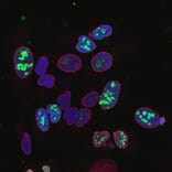
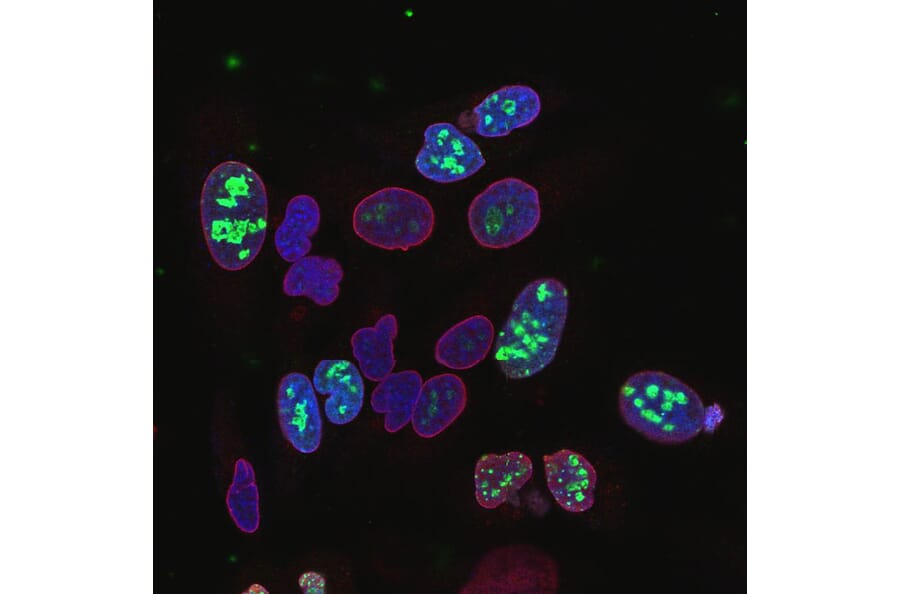
Immunofluorescence - Anti-Lamin A + Lamin C Antibody (A85443)
Confocal immunofluorescence image at high magnification of HeLa cell culture stained with Anti-Ki67 Antibody [6B4] (A104344), in green, and co-stained with Anti-Lamin A + Lamin C Antibody (A85443), in red. Anti-Ki67 Antibody [6B4] (A104344) stains the nuclei of rapidly dividing cells within their nucleoli, but doesn’t stain nearby quiescent cells. Anti-Lamin A + Lamin C Antibody (A85443) stains nuclear lamina. The blue is DAPI staining of nuclear DNA.
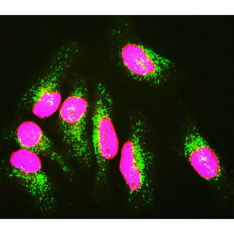
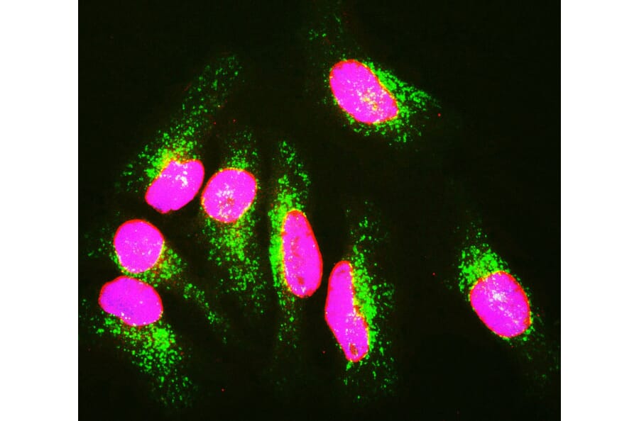
Immunofluorescence - Anti-Lamin A + Lamin C Antibody (A85443)
HeLa cells staining with Anti-Lamin A/C Antibody (red), Anti-Lamp1 Antibody (A85309 | green) and DAPI (DNA | blue). The Anti-Lamin A/C Antibody reveals strong nuclear lamina staining, while Anti-Lamp1 Antibody reveals strong cytoplasmic punctate staining of lysosomes and early endosomes. Since both DNA (blue) and Lamin A/C (red) are associated with the nuclear compartment, this region appears crimson in this image.
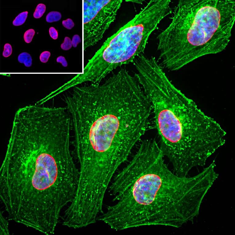
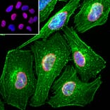
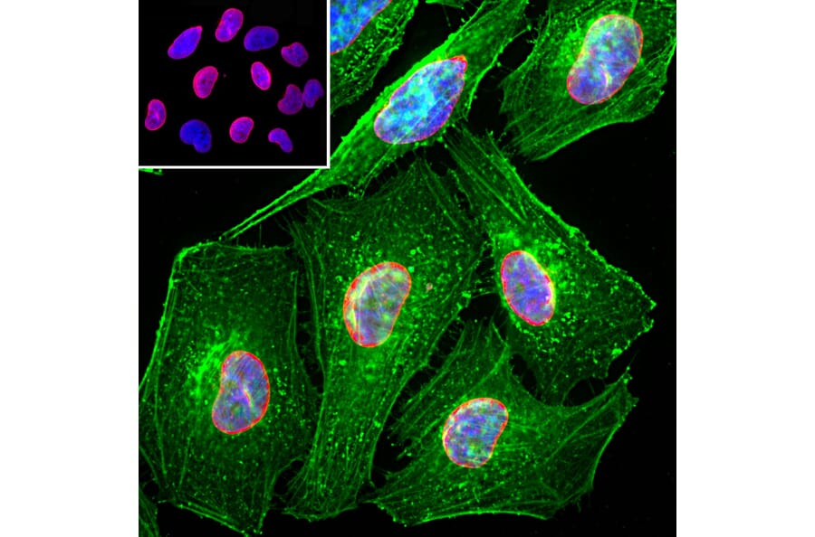
Immunofluorescence - Anti-Lamin A + Lamin C Antibody (A85443)
Immunofluorescent analysis of HeLa cells stained with Anti-Lamin A + C Antibody (A85443), at a dilution of 1:2,000 in red, and co-stained with Anti-Actin Antibody (A85388), at a dilution of 1:500, in green. The nuclear DNA is visualised in blue using Hoechst staining. The Anti-Lamin A + C Antibody (A85443) specifically labels the nuclear lamina, while the Anti-Actin Antibody (A85388) stains the submembranous actin-rich cytoskeleton, stress fibers and bundles of actin associated with cell adhesion sites.
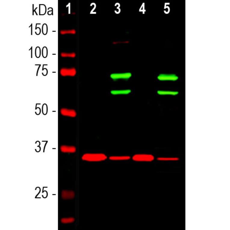
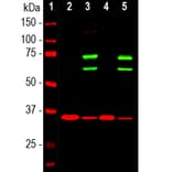
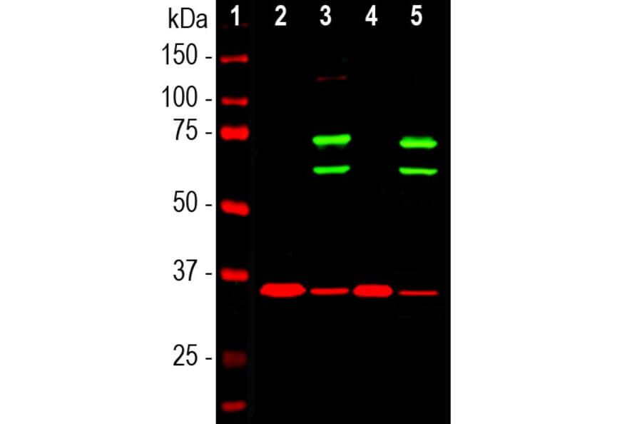
Western Blot - Anti-Lamin A + Lamin C Antibody (A85443)
Western blot analysis of cytosolic or nuclear enriched fractions of cell lines probed with Anti-Lamin A + C Antibody (A85443), at a dilution of 1:1,000, in green. The lanes contain samples of: [Lane 1] Protein standards, in red, [Lane 2] HeLa cytosol, [Lane 3] HeLa nuclear, [Lane 4] NIH-3T3 cytosol, and [Lane 5] NIH-3T3 nuclear fractions. Two strong bands at 65 kDa and 74 kDa correspond to Lamin A and Lamin C proteins respectively, detected exclusively in the nuclear fractions. The same blot was simultaneously probed with Anti-GAPDH Antibody (A85382), in red. The single band at 37 kDa represents GAPDH protein which is expressed predominantly in the cytosolic fractions.
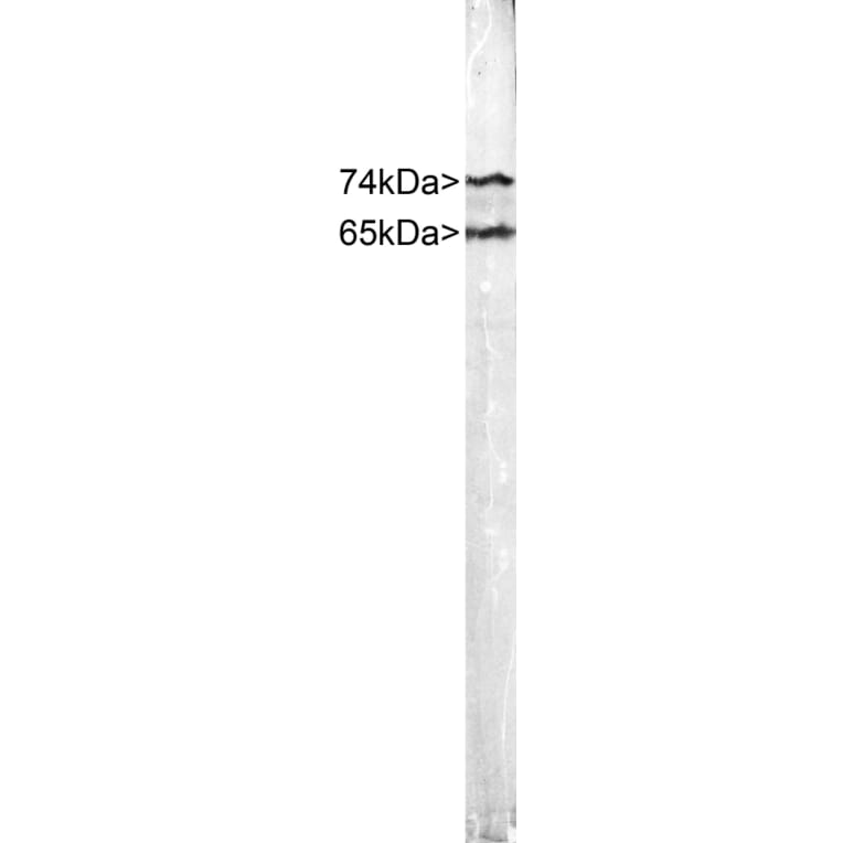
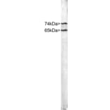
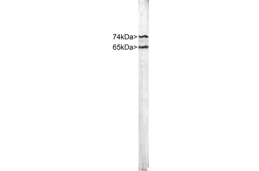
Western Blot - Anti-Lamin A + Lamin C Antibody (A85443)
Strip blot of crude HeLa cell extract stained with Anti-Lamin A/C Antibody. Note two strong clean bands at 74 kDa and 65 kDa, corresponding to Lamins A and C.
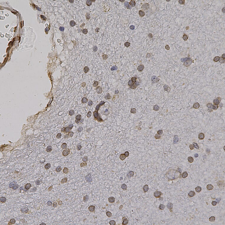
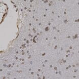
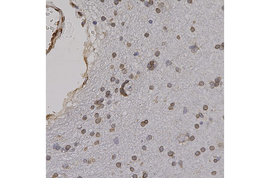
Immunohistochemistry - Anti-Lamin A + Lamin C Antibody (A85443)
Immunohistochemistry analysis of a formalin fixed paraffin embedded human cerebral cortex section with Anti-Lamin A + Lamin C Antibody (A85443) at a dilution of 1:2,000 detected in DAB (brown) following the ABC method. Counterstained with Hematoxylin (blue). Anti-Lamin A + Lamin C Antibody (A85443) recognizes the nuclear lamina, resulting in a distinct ring pattern.