Recombinant construct containing amino acids 1,111-1,490 of human Ki67 isotype 1, the central region of both Ki67 isotypes including the 2nd, 3rd, and 4th Ki67 repeat domains, expressed in and purified from E. coli.
Host
Rabbit
Clonality
Polyclonal
Isotype
IgG
Conjugate
Unconjugated
Molecular Weight
345 kDa, 395 kDa
Purity
Whole antiserum.
Product Form
Liquid
Formulation
Supplied as an aliquot of serum with 5mM Sodium Azide.
Storage
Shipped at 4°C. Upon delivery aliquot and store at -20°C. Avoid freeze/thaw cycles.
General Notes
Please note, Ki67 proteins are very unstable and only expressed in large amounts in situations where many cells are dividing. As a result of the very short half life of Ki67 there are usually numerous fragments visible on western blots running below the major 395 kDa and 345 kDa bands.
Synonyms
Antigen identified by monoclonal antibody Ki-67, Antigen KI-67, Antigen Ki67, MKI67, Proliferation marker protein Ki-67
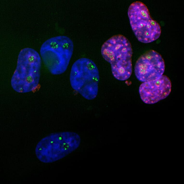
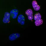
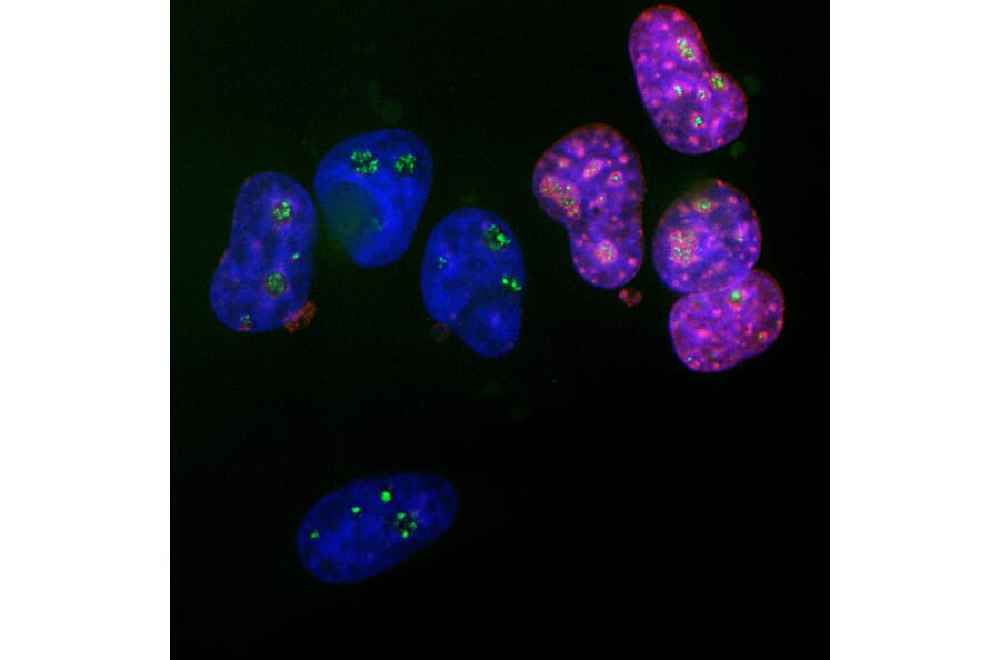
Immunofluorescent analysis of HeLa cells stained with Anti-Ki67 Antibody (A104333), at a dilution of 1:5,000, in red, and Anti-Fibrillarin Antibody [38F3] (A85370), at a dilution of 1:2,000, in green. The blue is DAPI staining of nuclear DNA. The Ki67 protein accumulates in and around the nucleoli of interphase cells such as those on the right, and the nucleoli are revealed by Anti-Fibrillarin Antibody [38F3] (A85370). In contrast, cells in the quiescent G0 state such as those on the left are Ki67 negative but Fibrillarin positive.
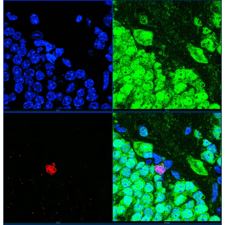
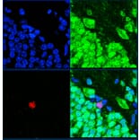
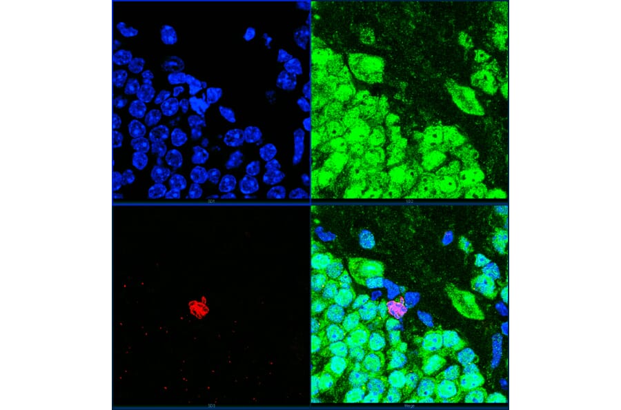
High magnification confocal image of adult mouse hippocampus dentate region stained with Anti-Ki67 Antibody (A104333) at a dilution of 1:2,000 (red) and Anti-NeuN Antibody [1B7] (A85405) at a dilution of 1:2,000 (green). Nuclei were stained with Hoechst (blue). Top left is DNA, top right FOX3/NeuN, bottom left Ki67 and bottom right all three merged. Dividing cells are very rare in adult animals, but one can be seen in the center of the image. Chromosomes can be seen in blue and their Ki67 coating can be seen (red). The dividing cell is FOX3/NeuN negative and so is presumably a glial cell.
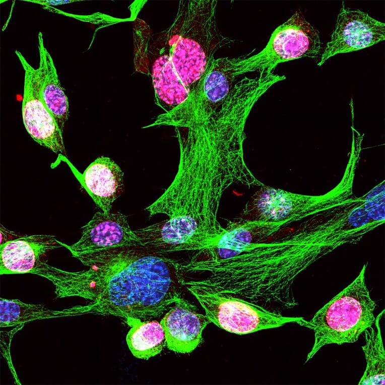
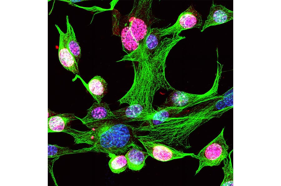
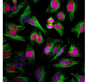
Mouse NIH-3T3 cells stained with Anti-Ki67 Antibody (A104333) at a dilution of 1:5,000 (red) and Anti-beta Tubulin Antibody [1B12] at a dilution of 1:2,000 (green). The Ki67 strongly stains the nuclei of dividing cells, but not quiescent cells. Nuclei were stained with Hoechst (blue).
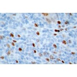
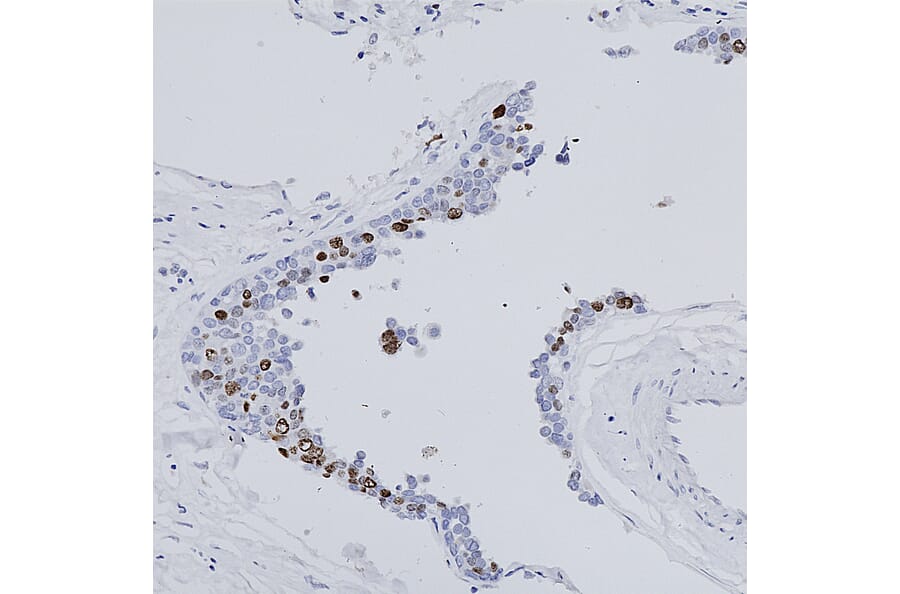
Immunohistochemistry analysis of formalin fixed paraffin embedded human breast tissue including normal and cancer cells. Cancer cells divide rapidly and heavily express Ki67 and so stain strongly with the Anti-Ki67 Antibody (A104333).
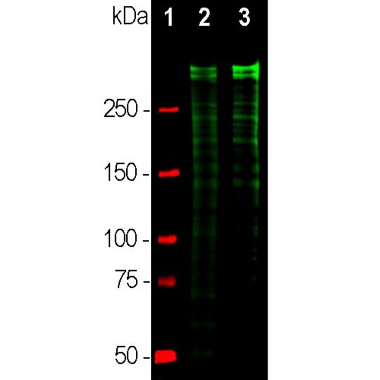
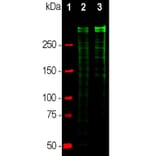
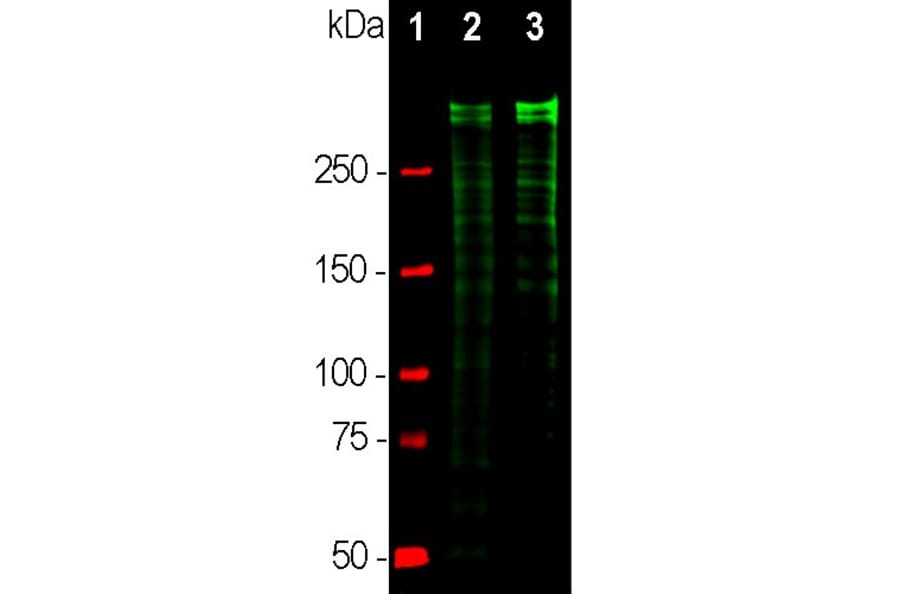
Western blot analysis of equal amounts of cell lysates using Anti-Ki67 Antibody (A104333), at a dilution of 1:10,000, in green. The lanes contain: [Lane 1] protein standard (red), [Lane 2] rapidly growing HeLa cell cultures, and [Lane 3] rapidly growing HEK293 cell cultures. Strong double bands larger than the 250kDa standard correspond to full length 345kDa and 395kDa Ki67 isoforms, while smaller proteolytic fragments of these isoforms are also invariably detected on the blot.
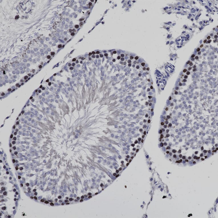
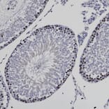
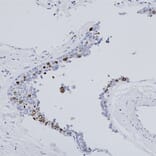
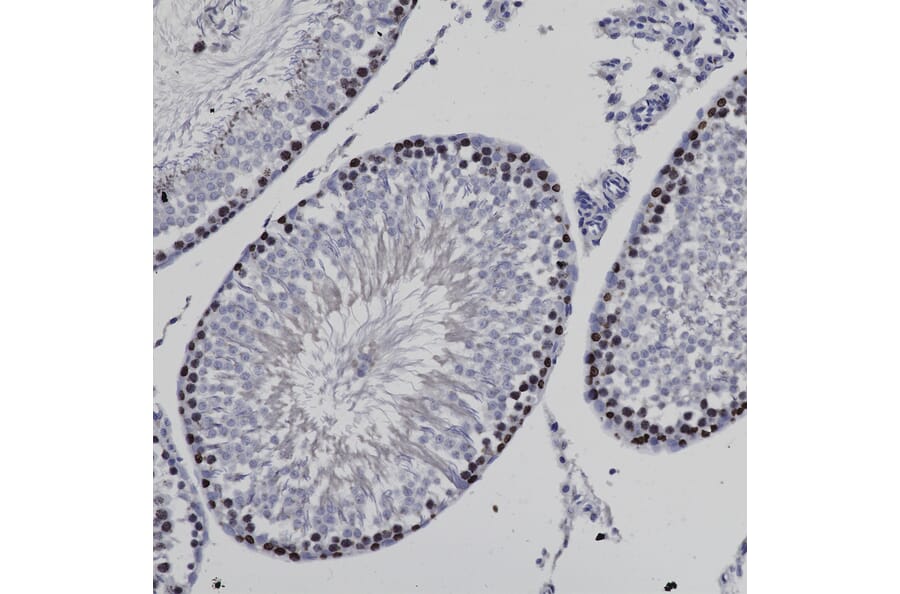
Immunohistochemistry analysis of a 4%PFA fixed paraffin embedded rat testes section with Anti-Ki67 Antibody (A104333) at a dilution of 1:5,000 detected with DAB (brown) using the Vector Labs ImmPRESS method and reagents with citra buffer retrieval. Counterstained with Hematoxylin (blue). In testes, the Anti-Ki67 Antibody (A104333) antibody strongly labels the nuclei of spermatogenic cells. Note: this antibody performs well in 4%PFA or NBF fixed tissues. Both rodent and human tissues stain effectively with Anti-Ki67 Antibody (A104333).
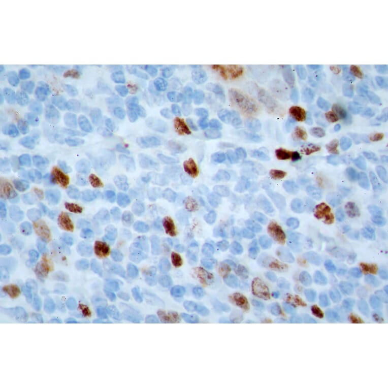
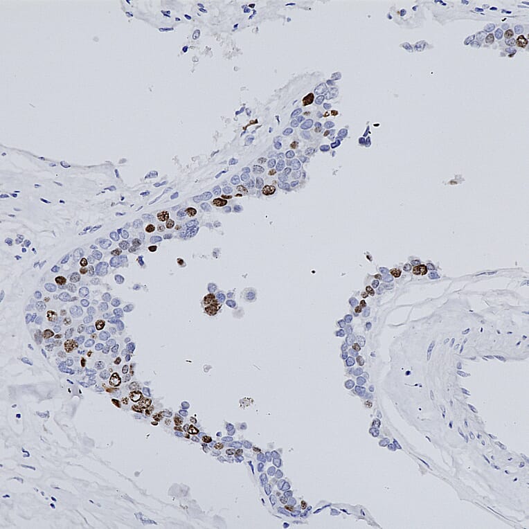
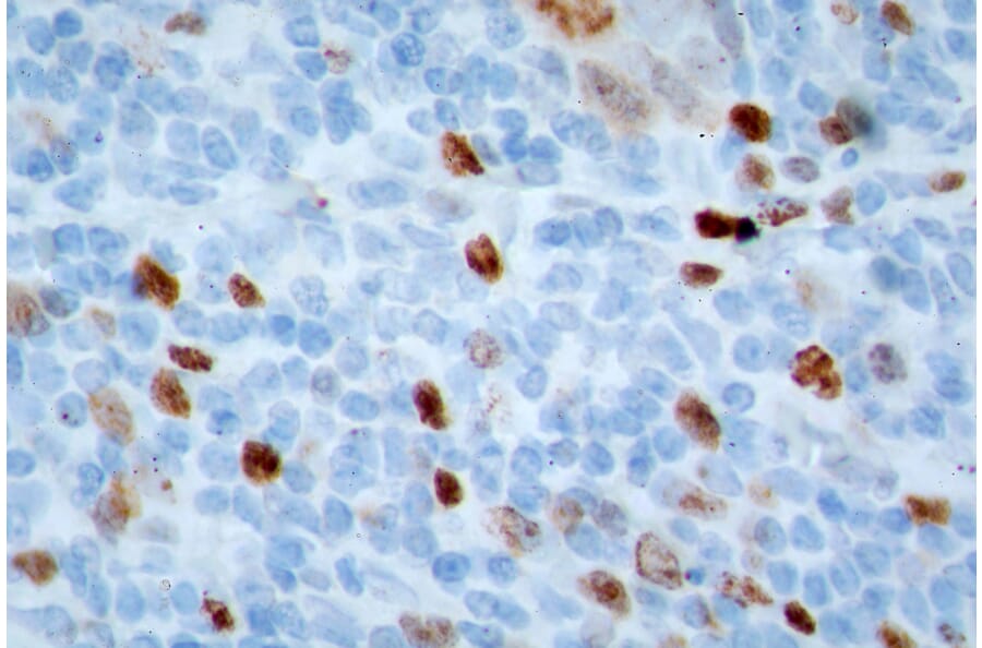
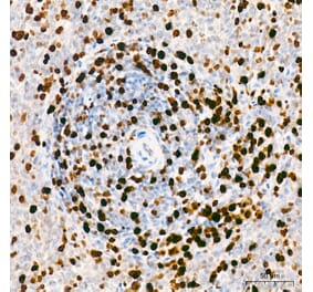
Immunohistochemistry analysis of a NBF fixed paraffin embedded human midbrain section with Anti-Ki67 Antibody (A104333) at a dilution of 1:5,000 detected with DAB (brown) using the Vector Labs ImmPRESS method and reagents with citra buffer retrieval. Counterstained with Hematoxylin (blue). Anti-Ki67 Antibody (A104333) strongly labels the nuclei of the actively dividing cancer cells.


![Immunocytochemistry - Anti-Ki67 Antibody [Ki-67] (A86642) - Antibodies.com](https://cdn.antibodies.com/image/catalog/86/A86644_785.jpg?profile=product_alternative)
![Immunohistochemistry - Anti-Ki67 Antibody [ARC5050-01] (A329549) - Antibodies.com](https://cdn.antibodies.com/image/catalog/329/A329549_1.jpg?profile=product_alternative)
![Immunohistochemistry - Anti-Ki67 Antibody [MKI67/2462] - BSA and Azide free (A252525) - Antibodies.com](https://cdn.antibodies.com/image/catalog/252/A252525_1.jpg?profile=product_alternative)
![Immunohistochemistry - Anti-Ki67 Antibody [MKI67/2462] (A249345) - Antibodies.com](https://cdn.antibodies.com/image/catalog/249/A249345_1.jpg?profile=product_alternative)
![Immunohistochemistry - Anti-Ki67 Antibody [MKI67/2461] - BSA and Azide free (A252522) - Antibodies.com](https://cdn.antibodies.com/image/catalog/252/A252522_1.jpg?profile=product_alternative)
![Immunohistochemistry - Anti-Ki67 Antibody [IHC067] (A86899) - Antibodies.com](https://cdn.antibodies.com/image/catalog/86/A86899_1.jpg?profile=product_alternative)
![Immunohistochemistry - Anti-Ki67 Antibody [MKI67/2461] (A249342) - Antibodies.com](https://cdn.antibodies.com/image/catalog/249/A249342_1.jpg?profile=product_alternative)
![Immunohistochemistry - Anti-Ki67 Antibody [MKI67/2465] (A249347) - Antibodies.com](https://cdn.antibodies.com/image/catalog/249/A249347_1.jpg?profile=product_alternative)
![Immunohistochemistry - Anti-Ki67 Antibody [MKI67/4945R] - BSA and Azide free (A252528) - Antibodies.com](https://cdn.antibodies.com/image/catalog/252/A252529_1.jpg?profile=product_alternative)
![Immunofluorescence - Anti-Ki67 Antibody [5F86] (A270555) - Antibodies.com](https://cdn.antibodies.com/image/catalog/270/A270555_1.jpg?profile=product_alternative)
![Immunohistochemistry - Anti-Ki67 Antibody [MKI67/2463] (A249346) - Antibodies.com](https://cdn.antibodies.com/image/catalog/249/A249346_1.jpg?profile=product_alternative)