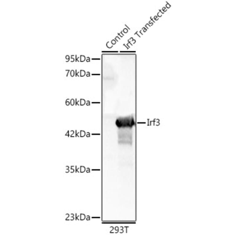
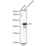
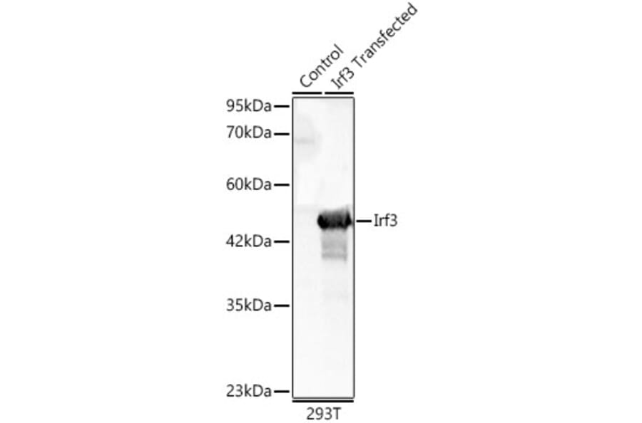
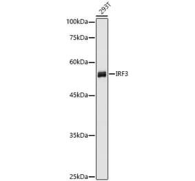
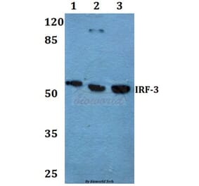
IRF3 expression in lysates from wild type (WT) and 293F cells transfected with Irf3 analyzed by western blot. Primary antibody incubation was performed for 1 hour on 25ug protein per lane with Anti-IRF3 Antibody (A329542) at a dilution of 1:900 and detected with chemiluminescence.
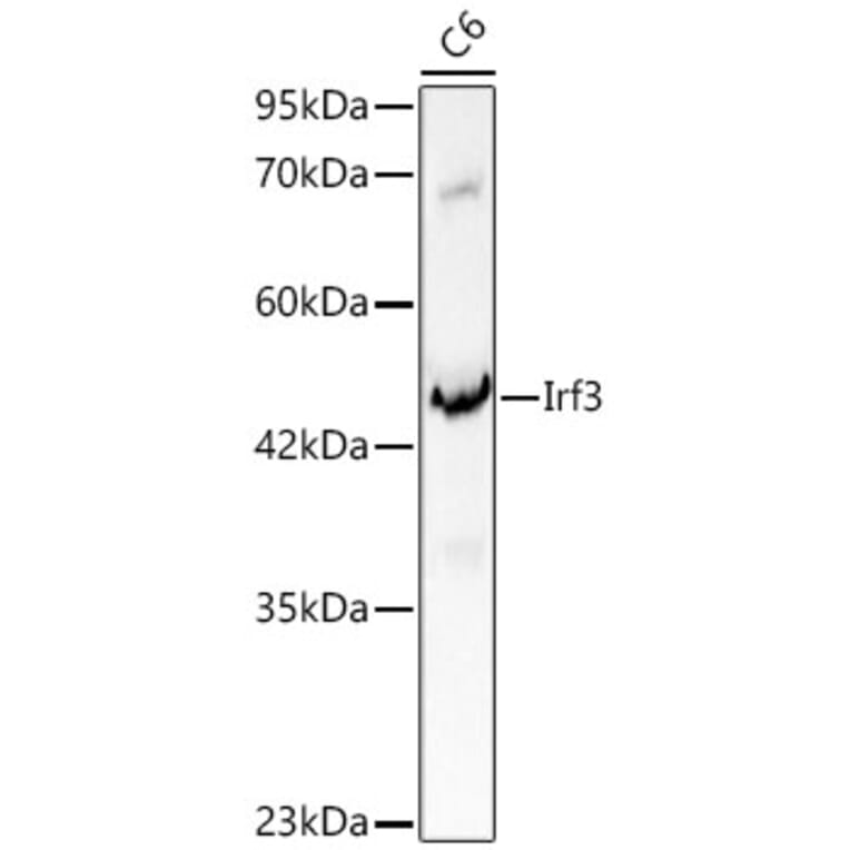
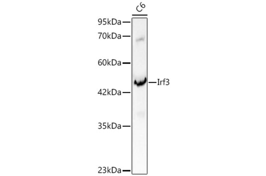
IRF3 expression in C6 cell lysates analyzed by western blot. Primary antibody incubation was performed for 1 hour on 25ug protein per lane with Anti-IRF3 Antibody (A329542) at a dilution of 1:900 and detected with chemiluminescence.
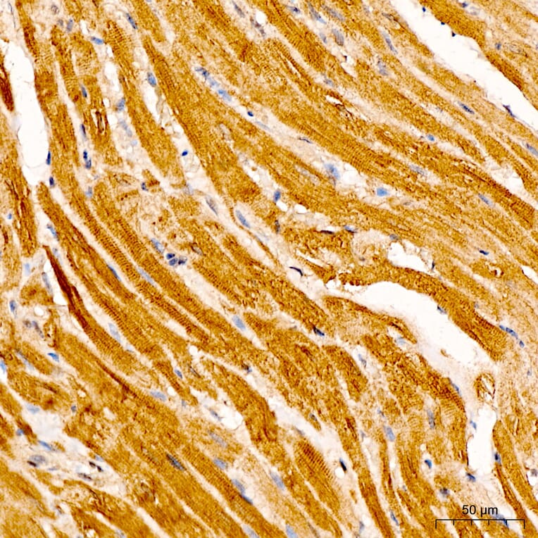
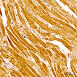
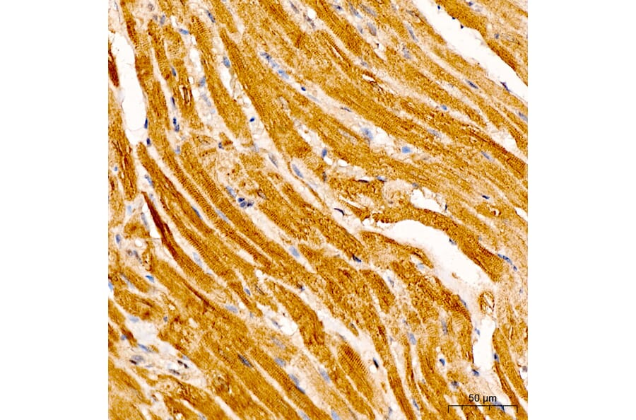
IRF3 expression in rat heart tissue analyzed by immunohistochemistry. Tissue was paraffin-embedded, and antigen retrieval was achieved with 10 mM citrate buffer, pH 6.0, under high pressure. Staining was performed with Anti-IRF3 Antibody (A329542) at a dilution of 1:100.
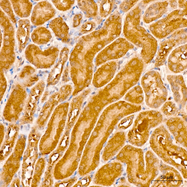
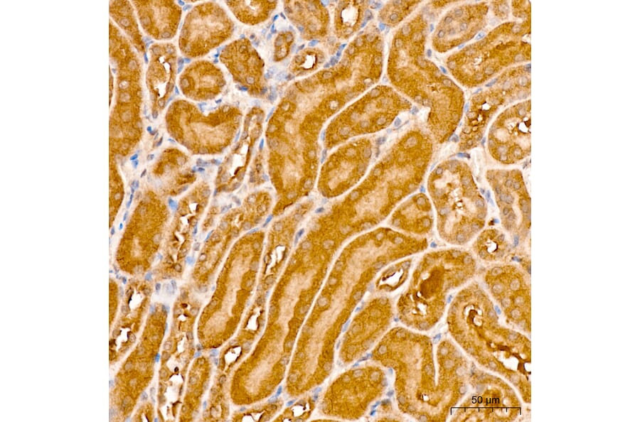
IRF3 expression in mouse kidney tissue analyzed by immunohistochemistry. Tissue was paraffin-embedded, and antigen retrieval was achieved with 10 mM citrate buffer, pH 6.0, under high pressure. Staining was performed with Anti-IRF3 Antibody (A329542) at a dilution of 1:100.
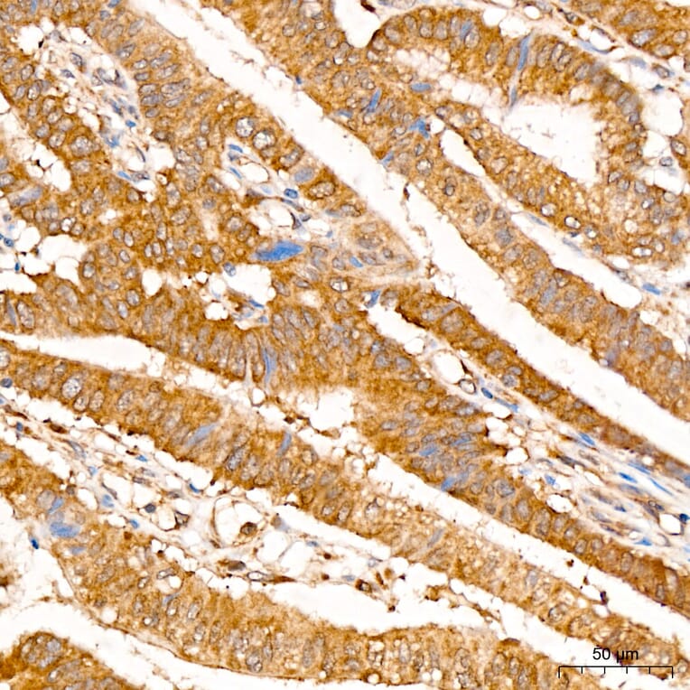
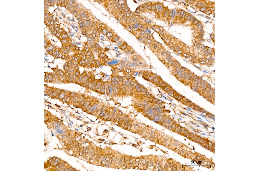
IRF3 expression in human colon carcinoma tissue analyzed by immunohistochemistry. Tissue was paraffin-embedded, and antigen retrieval was achieved with 10 mM citrate buffer, pH 6.0, under high pressure. Staining was performed with Anti-IRF3 Antibody (A329542) at a dilution of 1:100.


![Flow Cytometry - Anti-IRF3 Antibody [PCRP-IRF3-6C8] (A249050) - Antibodies.com](https://cdn.antibodies.com/image/catalog/249/A249050_1.jpg?profile=product_alternative)
![Flow Cytometry - Anti-IRF3 Antibody [PCRP-IRF3-6C8] - BSA and Azide free (A252230) - Antibodies.com](https://cdn.antibodies.com/image/catalog/252/A252230_1.jpg?profile=product_alternative)

![Immunofluorescence - Anti-IRF3 Antibody [PCRP-IRF3-1E6] (A249049) - Antibodies.com](https://cdn.antibodies.com/image/catalog/249/A249049_1.jpg?profile=product_alternative)
![Immunofluorescence - Anti-IRF3 Antibody [PCRP-IRF3-1E6] - BSA and Azide free (A252229) - Antibodies.com](https://cdn.antibodies.com/image/catalog/252/A252229_1.jpg?profile=product_alternative)
![SDS-PAGE - Anti-IRF3 Antibody [PCRP-IRF3-3B2] (A277870) - Antibodies.com](https://cdn.antibodies.com/image/catalog/277/A277870_1.jpg?profile=product_alternative)