Spontaneous autoantibody which binds LNERLAKLSDGVAVLKVGGT, amino acids 390-409 of the human sequence.
Host
Mouse
Clonality
Monoclonal
Clone ID
1C7
Isotype
IgG1
Conjugate
Unconjugated
Purification
Immunogen affinity purification.
Concentration
1 mg/ml
Molecular Weight
60 kDa
Product Form
Liquid
Formulation
Supplied in Phosphate Buffered Saline with 50% Glycerol and 5mM Sodium Azide.
Storage
Shipped at 4°C. Upon delivery aliquot and store at -20°C. Avoid freeze/thaw cycles.
General Notes
This antibody was discovered during screens for antibodies produced by hybridomas from a mouse injected with an unrelated protein. We noted beautiful staining of mitochondria and clean staining of a single band of 60 kDa on western blots, suggesting HSP60 as an obvious candidate. We made recombinant full-length human HSP60 which this antibody bound to strongly and specifically. The mouse had likely developed autoantibodies to HSP60 since this molecule appears to be unusually immunogenic, frequently generating autoantibodies in humans and other species.
Synonyms
60 kDa chaperonin, 60 kDa heat shock protein, mitochondrial, Chaperonin 60, CPN60, Heat shock protein 60, Heat shock protein family D member 1, HSP-60, HSPD1, HuCHA60
Western blot analysis of tissue or whole cell lysates using Anti-Heat Shock Protein 60 Antibody, at a dilution of 1:10,000, in green,: [Lane 1] protein standard (red), [Lane 2] rat brain, [Lane 3] mouse brain, [Lane 4] NIH/3T3, [Lane 5] Hek 293, [Lane 6] HeLa, [Lane 7] SH-SY5Y cells. Strong single band corresponds to HSP60 protein with apparent SDS-PAGE molecular weight of 60 kDa.
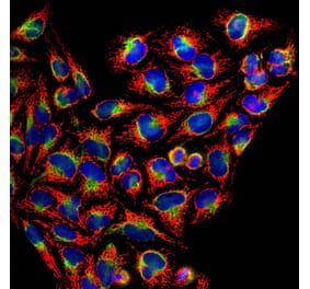
Immunofluorescent analysis of HeLa cells stained with Anti-Heat Shock Protein 60 Antibody [1C7] (A85439), at a dilution of 1:5,000, in red. Blue is DAPI staining of nuclear DNA. Anti-Heat Shock Protein 60 Antibody [1C7] (A85439) produces strong and specific staining of mitochondria.
Immunohistochemistry analysis of a 4% PFA fixed paraffin embedded rat cerebellum section with Anti-HSP60 Antibody [1C7] (A85439) at a dilution of 1:1,000 detected with DAB (brown) using the ImmPress method, with citra buffer retrieval. Counterstained with Hematoxylin (blue). Note: this antibody performs well in testing with both 4% PFA and standard NBF fixed tissues but does not stain long term NBF fixed tissue effectively.
Western blot analysis of tissue or whole cell lysates using Anti-HSP60 Antibody [1C7] (A85439) at a dilution of 1:10,000, (green). [1] protein standard (red), [Lane 2] rat brain, [Lane 3] mouse brain, [Lane 4] NIH-3T3, [Lane 5] HEK293, [Lane 6] HeLa, and [Lane 7] SH-SY5Y cells. Strong single band corresponds to HSP60 protein with apparent SDS-PAGE molecular weight of 60kDa.
Western blot analysis of Anti-Heat Shock Protein 60 Antibody. Blot of HeLa cells lysates was probed with Anti-Heat Shock Protein 60 Antibody. This antibody recognizes HSP60 at 60 kDa.


![Western Blot - Anti-HSP60 Antibody [1C7] (A85439) - Antibodies.com](https://cdn.antibodies.com/image/catalog/85/A85439_1.jpg?profile=product_top)
![Immunofluorescence - Anti-HSP60 Antibody [1C7] (A85439) - Antibodies.com](https://cdn.antibodies.com/image/catalog/85/A85439_2.jpg?profile=product_top)
![Immunohistochemistry - Anti-HSP60 Antibody [1C7] (A85439) - Antibodies.com](https://cdn.antibodies.com/image/catalog/85/A85439_3.jpg?profile=product_top)
![Western Blot - Anti-HSP60 Antibody [1C7] (A85439) - Antibodies.com](https://cdn.antibodies.com/image/catalog/85/A85439_4.jpg?profile=product_top)
![Western Blot - Anti-HSP60 Antibody [1C7] (A85439) - Antibodies.com](https://cdn.antibodies.com/image/catalog/85/A85439_5.jpg?profile=product_top)
![Western Blot - Anti-HSP60 Antibody [1C7] (A85439) - Antibodies.com](https://cdn.antibodies.com/image/catalog/85/A85439_1.jpg?profile=product_top_thumb)
![Immunofluorescence - Anti-HSP60 Antibody [1C7] (A85439) - Antibodies.com](https://cdn.antibodies.com/image/catalog/85/A85439_2.jpg?profile=product_top_thumb)
![Immunohistochemistry - Anti-HSP60 Antibody [1C7] (A85439) - Antibodies.com](https://cdn.antibodies.com/image/catalog/85/A85439_3.jpg?profile=product_top_thumb)
![Western Blot - Anti-HSP60 Antibody [1C7] (A85439) - Antibodies.com](https://cdn.antibodies.com/image/catalog/85/A85439_4.jpg?profile=product_top_thumb)
![Western Blot - Anti-HSP60 Antibody [1C7] (A85439) - Antibodies.com](https://cdn.antibodies.com/image/catalog/85/A85439_5.jpg?profile=product_top_thumb)
![Western Blot - Anti-HSP60 Antibody [1C7] (A85439) - Antibodies.com](https://cdn.antibodies.com/image/catalog/85/A85439_1.jpg?profile=product_image)
![Immunofluorescence - Anti-HSP60 Antibody [1C7] (A85439) - Antibodies.com](https://cdn.antibodies.com/image/catalog/85/A85439_2.jpg?profile=product_image)
![Immunohistochemistry - Anti-HSP60 Antibody [1C7] (A85439) - Antibodies.com](https://cdn.antibodies.com/image/catalog/85/A85439_3.jpg?profile=product_image)
![Western Blot - Anti-HSP60 Antibody [1C7] (A85439) - Antibodies.com](https://cdn.antibodies.com/image/catalog/85/A85439_4.jpg?profile=product_image)
![Western Blot - Anti-HSP60 Antibody [1C7] (A85439) - Antibodies.com](https://cdn.antibodies.com/image/catalog/85/A85439_5.jpg?profile=product_image)
![Immunocytochemistry/Immunofluorescence - Anti-HSP60 Antibody [LK1] (A305088) - Antibodies.com](https://cdn.antibodies.com/image/catalog/305/A305088_1.png?profile=product_alternative)
![Immunofluorescence - Anti-HSP60 Antibody [LK2] (A248884) - Antibodies.com](https://cdn.antibodies.com/image/catalog/248/A248885_1.jpg?profile=product_alternative)
![Immunofluorescence - Anti-HSP60 Antibody [LK2] - BSA and Azide free (A252064) - Antibodies.com](https://cdn.antibodies.com/image/catalog/252/A252065_1.jpg?profile=product_alternative)
![Immunohistochemistry - Anti-HSP60 Antibody [LK2] (A305090) - Antibodies.com](https://cdn.antibodies.com/image/catalog/305/A305090_1.png?profile=product_alternative)
![Immunohistochemistry - Anti-HSP60 Antibody [HSPD1/780] (A248887) - Antibodies.com](https://cdn.antibodies.com/image/catalog/248/A248887_1.jpg?profile=product_alternative)
![Immunohistochemistry - Anti-HSP60 Antibody [LK1] (A248883) - Antibodies.com](https://cdn.antibodies.com/image/catalog/248/A248883_1.jpg?profile=product_alternative)
![Immunohistochemistry - Anti-HSP60 Antibody [HSPD1/780] - BSA and Azide free (A252067) - Antibodies.com](https://cdn.antibodies.com/image/catalog/252/A252067_1.jpg?profile=product_alternative)
![Immunohistochemistry - Anti-HSP60 Antibody [LK1] - BSA and Azide free (A252063) - Antibodies.com](https://cdn.antibodies.com/image/catalog/252/A252063_1.jpg?profile=product_alternative)
![Immunohistochemistry - Anti-HSP60 Antibody [SPM253] (A248884) - Antibodies.com](https://cdn.antibodies.com/image/catalog/248/A248884_1.jpg?profile=product_alternative)
![Immunohistochemistry - Anti-HSP60 Antibody [SPM253] - BSA and Azide free (A252064) - Antibodies.com](https://cdn.antibodies.com/image/catalog/252/A252064_1.jpg?profile=product_alternative)