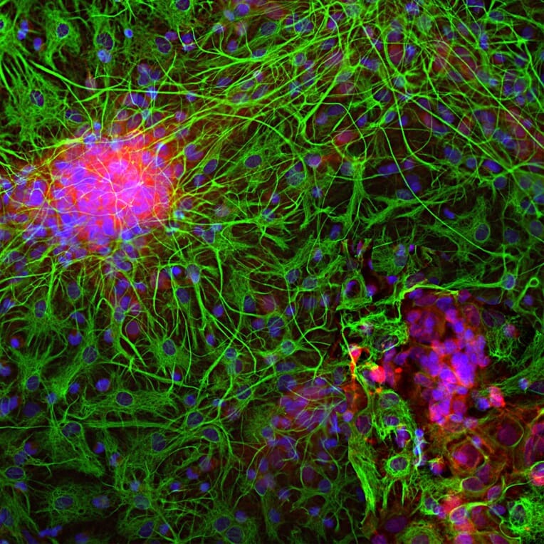
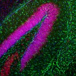
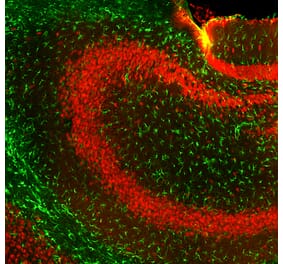
Immunofluorescent analysis of a section of mouse hippocampus stained with Anti-GFAP Antibody, at a dilution of 1:5,000, in green, and co-stained with Anti-FOX3/NeuN Antibody (A85402 | 1:5,000, in red. The blue is DAPI staining of nuclear DNA. Following transcardial perfusion with 4% paraformaldehyde, mouse brain was post fixed for 24 hours, cut to 45 µM, and free-floating sections were stained with the above antibodies. The Anti-GFAP Antibody stains a network of astroglial cells while the Anti-Fox3/NeuN Antibody stains the nuclei and proximal perikarya of neurons.
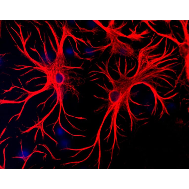
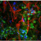
Mixed cultures of neurons and glia stained with Anti-GFAP Antibody (red) and DNA (blue). Astrocytes stain strongly and specifically in a clearly filamentous fashion with this antibody.
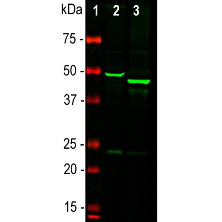
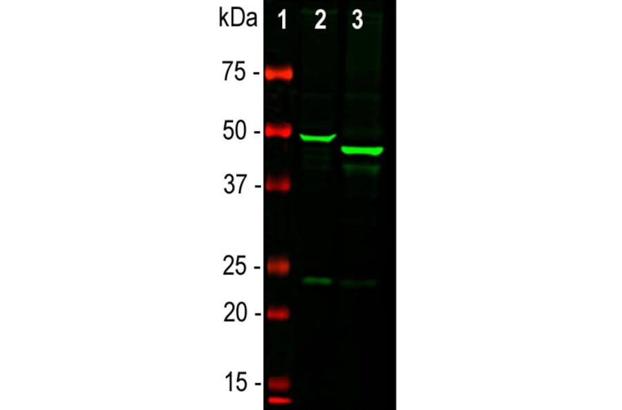
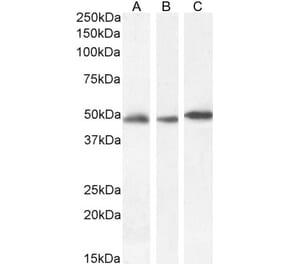
Western blot analysis of whole brain lysates using Anti-GFAP Antibody, at a dilution of 1:5,000, in green,: [Lane 1] protein standard (red), [Lane 2] rat brain, [Lane 3] mouse brain. The strong band at about 50 kDa corresponds to the GFAP protein. Smaller proteolytic fragments and alternate transcripts of GFAP may also be detected on such blots.
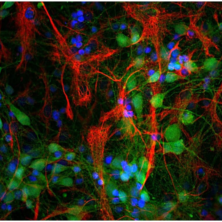
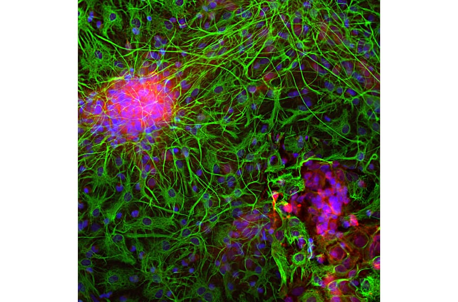
Immunofluorescent analysis of mixed cortical neuron-glial cell culture from E20 rat stained with Anti-NSE Antibody (A85285), at a dilution of 1:500, in red, and co-stained with Anti-GFAP Antibody (A85307), at a dilution of 1:5,000 in green. The nuclear DNA is visualised in blue using Hoechst staining. Anti-NSE Antibody (A85285) labels protein expressed in neuronal cells, while the Anti-GFAP Antibody (A85307) stains intermediate filaments in astrocytic and certain other glial cells.
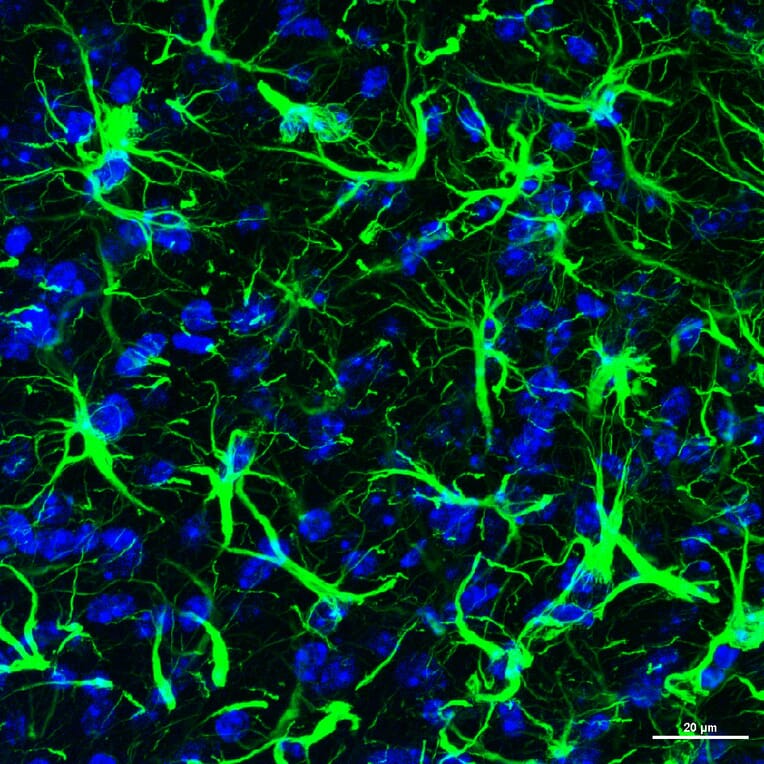
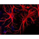
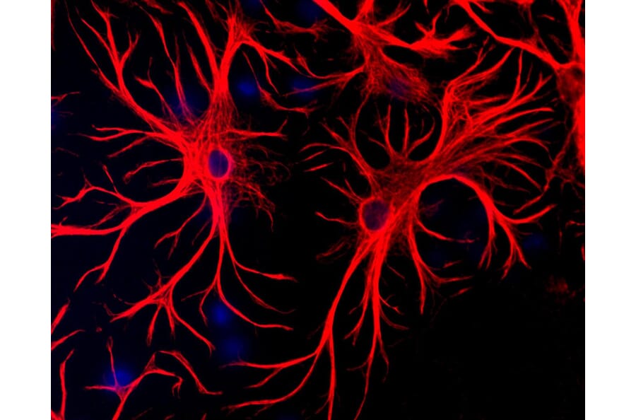
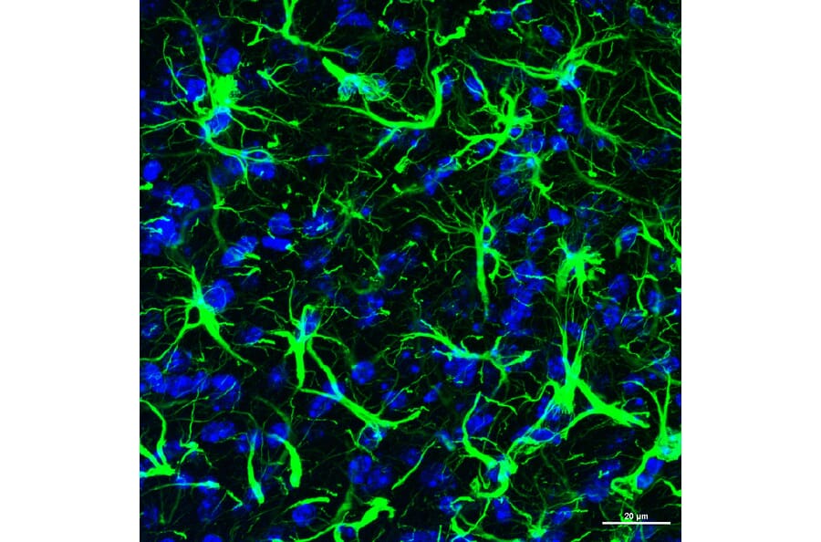
A section of mouse frontal cerebral cortex was stained with Anti-GFAP Antibody (A85307) (green) and nuclei were stained with DAPI (blue). The fibrous component of the processes of astroctyes are clearly revealed.
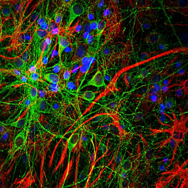
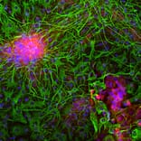
Immunofluorescent analysis of cortical neuron-glial cell culture from E20 rat costained with Anti-GFAP Antibody (A85307) at a dilution of 1:2,000 (red) and Anti-Tau Antibody [5B10] (A85415) at a dilution of 1:2,000 (green). Nuclei were stained with DAPI (blue). The GFAP labels astroglial cells, while the MAP-t antibody stains neuronal cell perikarya, dendrites and axons.
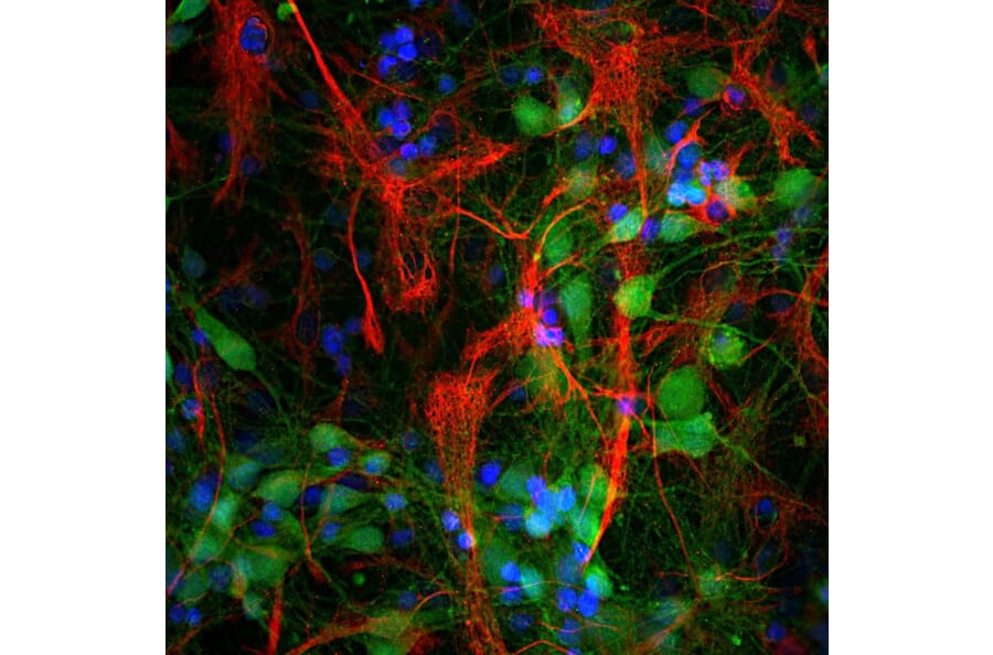
Immunofluorescent analysis of cortical neuron-glial culture from E20 rat stained with Anti-UCHL1 Antibody [BH7] (A85351) at a dilution of 1:5,000 (green) and costained with Anti-GFAP Antibody (A85307) at a dilution of 1:5,000 (red). Nuclei were stained with DAPI (blue). The Anti-UCHL1 Antibody [BH7] (A85351) antibody stains cell bodies and dendrites of neurons, while the GFAP antibody labels astrocytes.
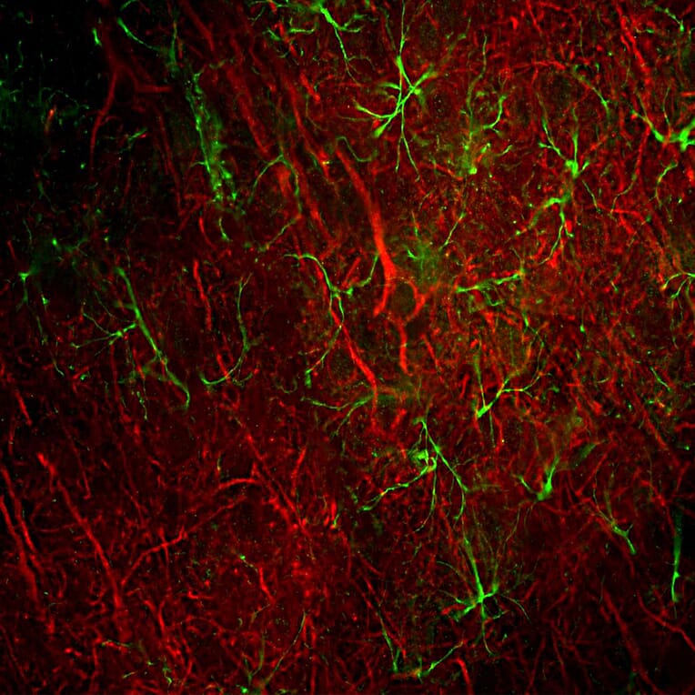
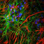
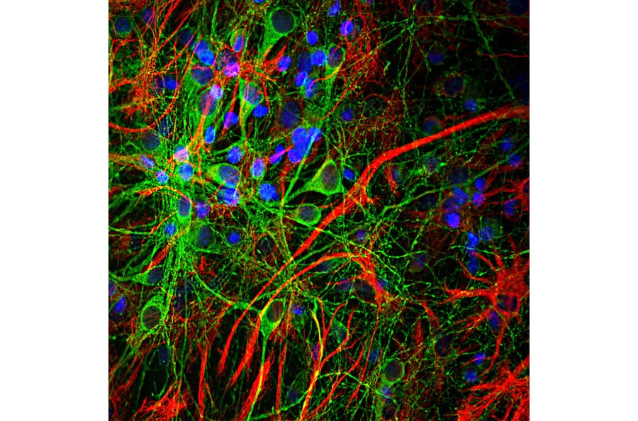
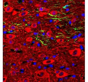
Immunofluorescent analysis of rat frontal cortex section stained with Anti-NF-L Antibody [DA2] (A85454) at a dilution of 1:500 (red) and costained with Anti-GFAP Antibody (A85307) at a dilution of 1:5,000 (green). Following transcardial perfusion of rat with 4% paraformaldehyde, brain was post fixed for 24 hours, cut to 45µM, and free-floating sections were stained with above antibodies. This antibody labels cell bodies and processes of pyramidal neurons, as well as dendrites and axons of other neuronal cells, while the GFAP antibody stains the network of glial cells.
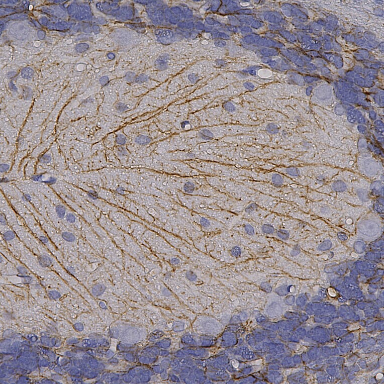
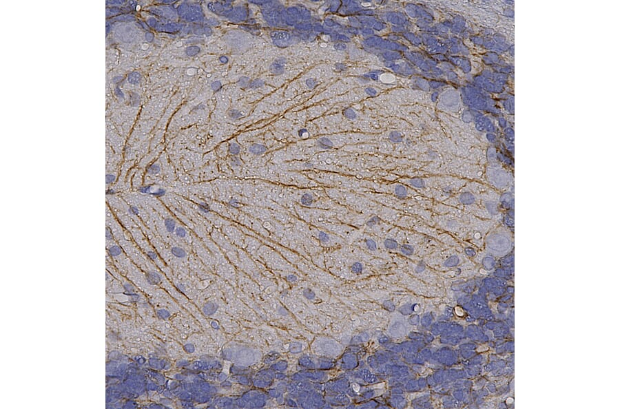
Immunohistochemistry analysis of a formalin fixed paraffin embedded human cerebellum section with Anti-GFAP Antibody (A85307) at a dilution of 1:20,000 detected in DAB (brown) following the ABC method. Counterstained with Hematoxylin (blue). The GFAP antibody detects the core of processes of astrocytes and Bergman glia within the granular and molecular layers.


![Immunofluorescence - Anti-GFAP Antibody [2A5] (A104314) - Antibodies.com](https://cdn.antibodies.com/image/catalog/104/A104314_1.jpg?profile=product_alternative)
![Immunofluorescence - Anti-GFAP Antibody [5C10] (A85422) - Antibodies.com](https://cdn.antibodies.com/image/catalog/85/A85422_1.jpg?profile=product_alternative)
![Western Blot - Anti-GFAP Antibody [GA-5] - BSA and Azide free (A251887) - Antibodies.com](https://cdn.antibodies.com/image/catalog/251/A251887_1.jpg?profile=product_alternative)
![Western Blot - Anti-GFAP Antibody [GA-5] (A248705) - Antibodies.com](https://cdn.antibodies.com/image/catalog/248/A248705_1.jpg?profile=product_alternative)
![Immunohistochemistry - Anti-GFAP Antibody [SPM507] - BSA and Azide free (A251888) - Antibodies.com](https://cdn.antibodies.com/image/catalog/251/A251889_1.jpg?profile=product_alternative)
![Immunohistochemistry - Anti-GFAP Antibody [GA-5 + ASTRO/789] (A248708) - Antibodies.com](https://cdn.antibodies.com/image/catalog/248/A248709_1.jpg?profile=product_alternative)
![Immunohistochemistry - Anti-GFAP Antibody [SPM248] - BSA and Azide free (A251888) - Antibodies.com](https://cdn.antibodies.com/image/catalog/251/A251888_1.jpg?profile=product_alternative)
![Immunohistochemistry - Anti-GFAP Antibody [GA-5 + ASTRO/789] - BSA and Azide free (A251890) - Antibodies.com](https://cdn.antibodies.com/image/catalog/251/A251891_1.jpg?profile=product_alternative)

![Western Blot - Anti-GFAP Antibody [ARC0206] (A307282) - Antibodies.com](https://cdn.antibodies.com/image/catalog/307/A307282_1.jpg?profile=product_alternative)
![Immunohistochemistry - Anti-GFAP Antibody [SPM248] (A248706) - Antibodies.com](https://cdn.antibodies.com/image/catalog/248/A248706_1.jpg?profile=product_alternative)