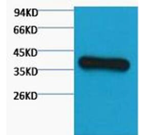
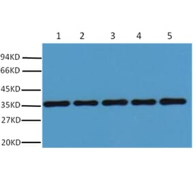
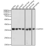
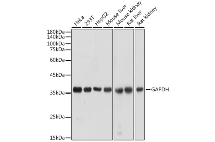
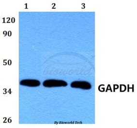
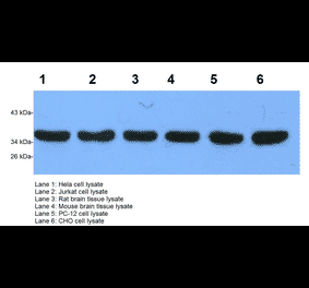
Western blot analysis of extracts of various cell lines, using Anti-GAPDH Antibody (A92899) at 1:10,000 dilution. Lysates/proteins were present at 25µg per lane. The blocking buffer used was 3% non-fat dry milk in TBST. Detection was with a ECL Basic Kit. Exposure time: 1s.
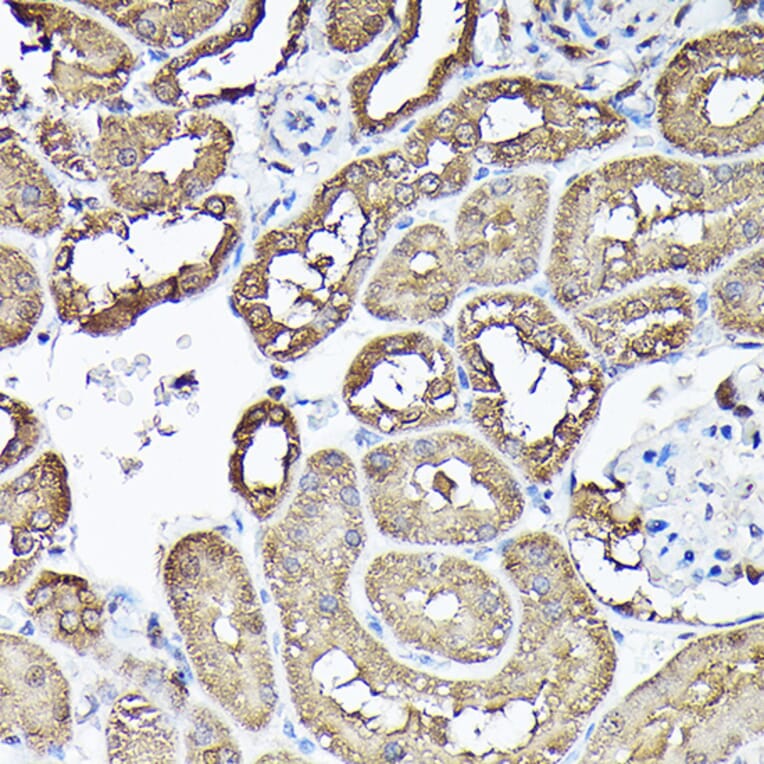
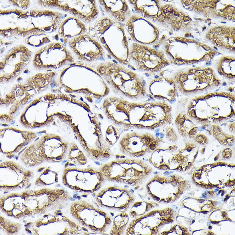
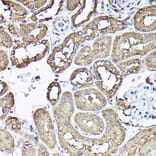
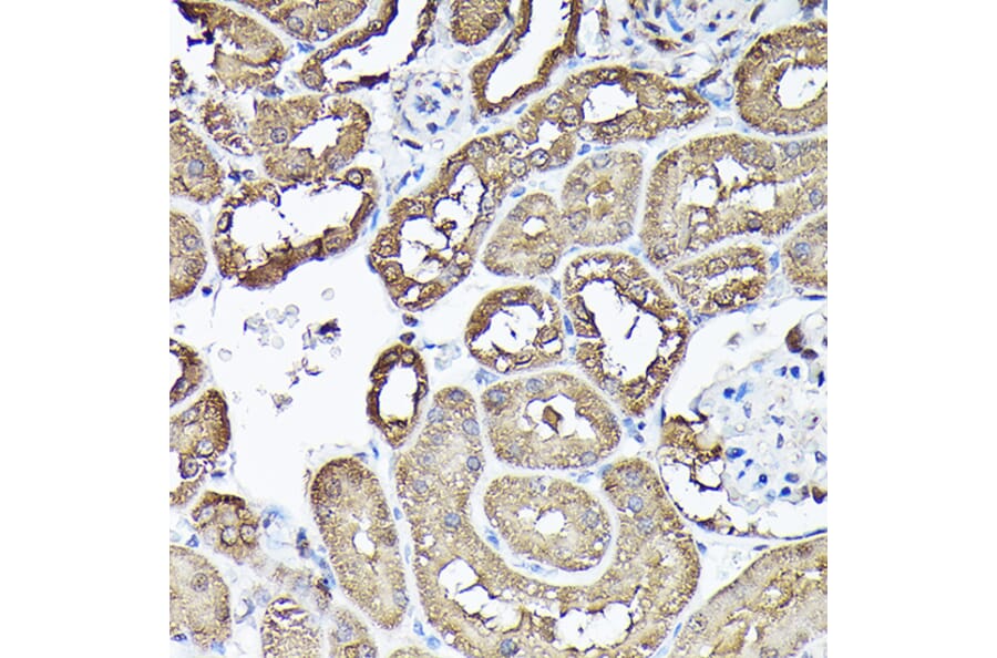
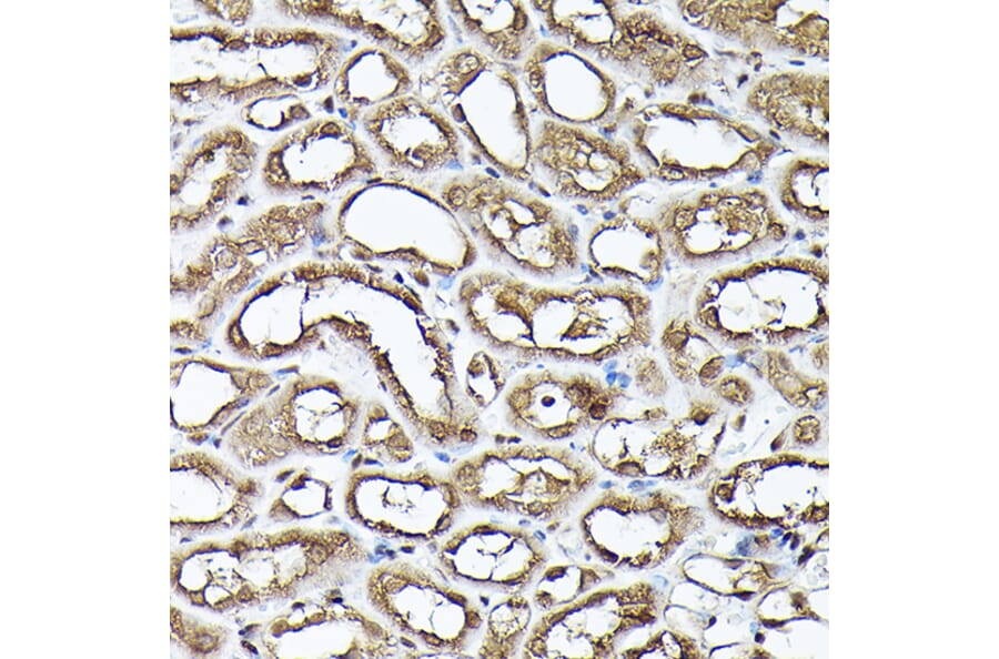
Immunohistochemistry analysis of paraffin-embedded rat kidney using Anti-GAPDH Antibody (A92899)Immunohistochemistry analysis of paraffin-embedded rat kidney using GAPDH Rabbit polyclonal antibody (AC027) at a dilution of 1:100 (40x lens). Perform high pressure antigen retrieval with 10 mM citrate buffer pH 6.0 before commencing with IHC staining protocol.
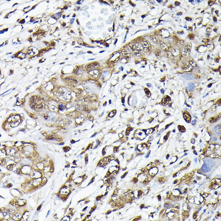
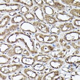
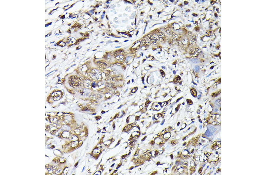
Immunohistochemistry analysis of paraffin-embedded human liver cancer using Anti-GAPDH Antibody (A92899)Immunohistochemistry analysis of paraffin-embedded human liver cancer using GAPDH Rabbit polyclonal antibody (AC027) at a dilution of 1:100 (40x lens). Perform high pressure antigen retrieval with 10 mM citrate buffer pH 6.0 before commencing with IHC staining protocol.
Immunohistochemistry analysis of paraffin-embedded mouse kidney using Anti-GAPDH Antibody (A92899)Immunohistochemistry analysis of paraffin-embedded mouse kidney using GAPDH Rabbit polyclonal antibody (AC027) at a dilution of 1:100 (40x lens). Perform high pressure antigen retrieval with 10 mM citrate buffer pH 6.0 before commencing with IHC staining protocol.
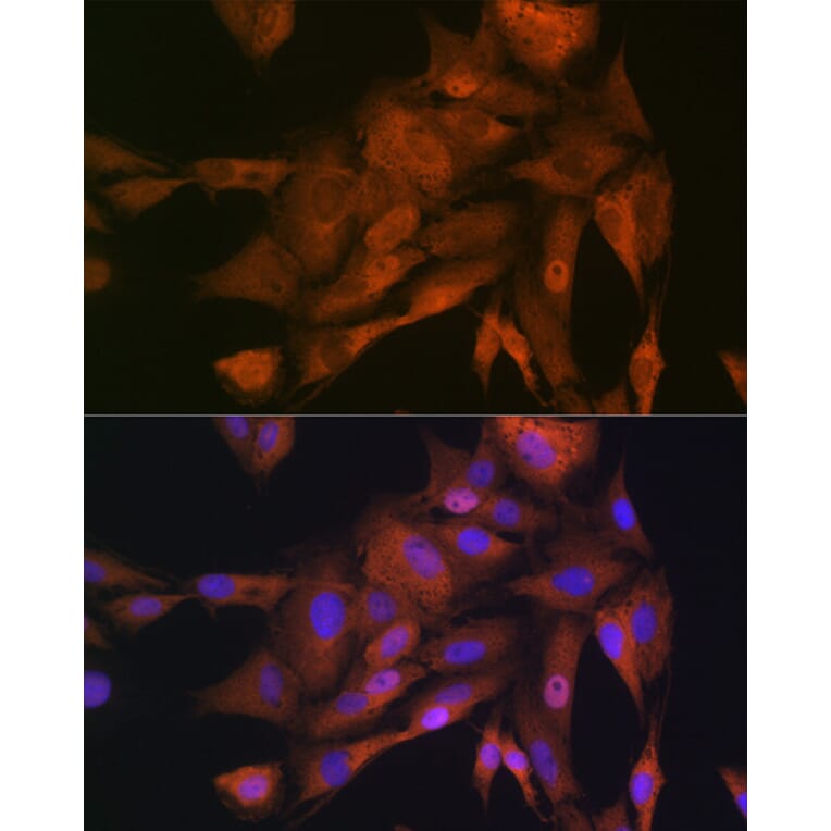
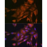
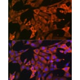
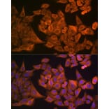
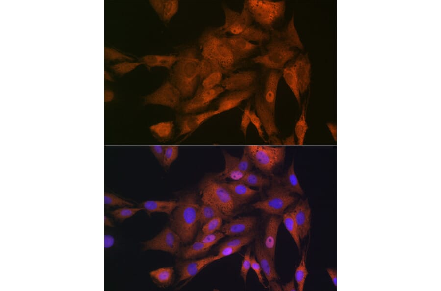
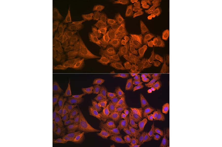
Immunofluorescence analysis of C6 cells using Anti-GAPDH Antibody (A92899) at a dilution of 1:100 (40x lens). DAPI was used to stain the cell nuclei (blue).
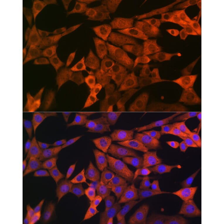
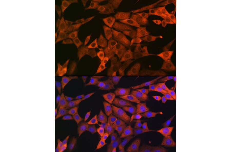
Immunofluorescence analysis of NIH-3T3 cells using Anti-GAPDH Antibody (A92899) at a dilution of 1:100 (40x lens). DAPI was used to stain the cell nuclei (blue).
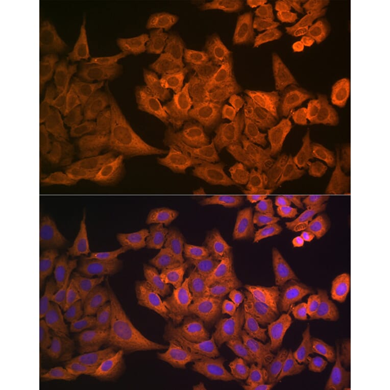
Immunofluorescence analysis of U-2 OS cells using Anti-GAPDH Antibody (A92899) at a dilution of 1:100 (40x lens). DAPI was used to stain the cell nuclei (blue).


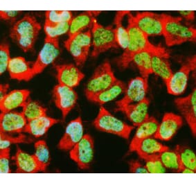
![Immunofluorescence - Anti-GAPDH Antibody [1D4] (A85382) - Antibodies.com](https://cdn.antibodies.com/image/catalog/85/A85382_1.jpg?profile=product_alternative)
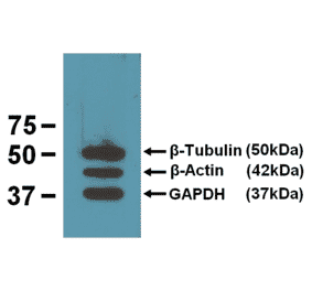

![Western Blot - Anti-GAPDH Antibody [ARC50888] (A309068) - Antibodies.com](https://cdn.antibodies.com/image/catalog/309/A309068_1.jpg?profile=product_alternative)
![Immunocytochemistry - Anti-GAPDH Antibody [RM114] (A121303) - Antibodies.com](https://cdn.antibodies.com/image/catalog/121/A121396_1.png?profile=product_alternative)