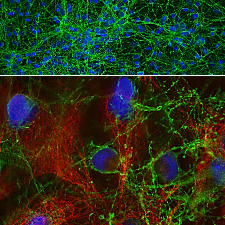
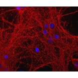
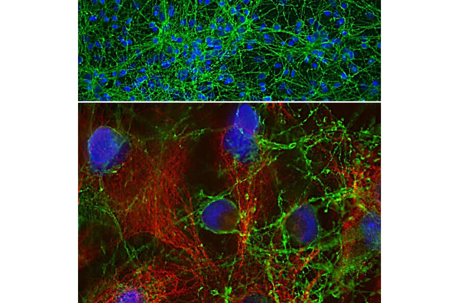
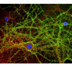
Immunofluorescent analysis of cortical neuron-glial cell culture from E20 rat stained with Anti-GAP43 Antibody, at a dilution of 1:2,000, in green, and Anti-Vimentin Antibody (A85423 | 1:2,000, in red. The blue is DAPI staining of nuclear DNA. Anti-GAP43 Antibody labels protein expressed in the axonal membrane of neuronal cells, while Anti-Vimentin Antibody stains intermediate filaments in fibroblasts and other non-neuronal cells.
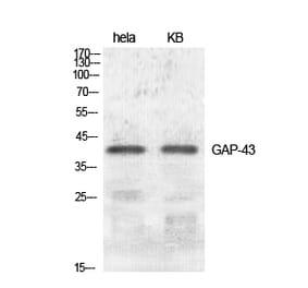
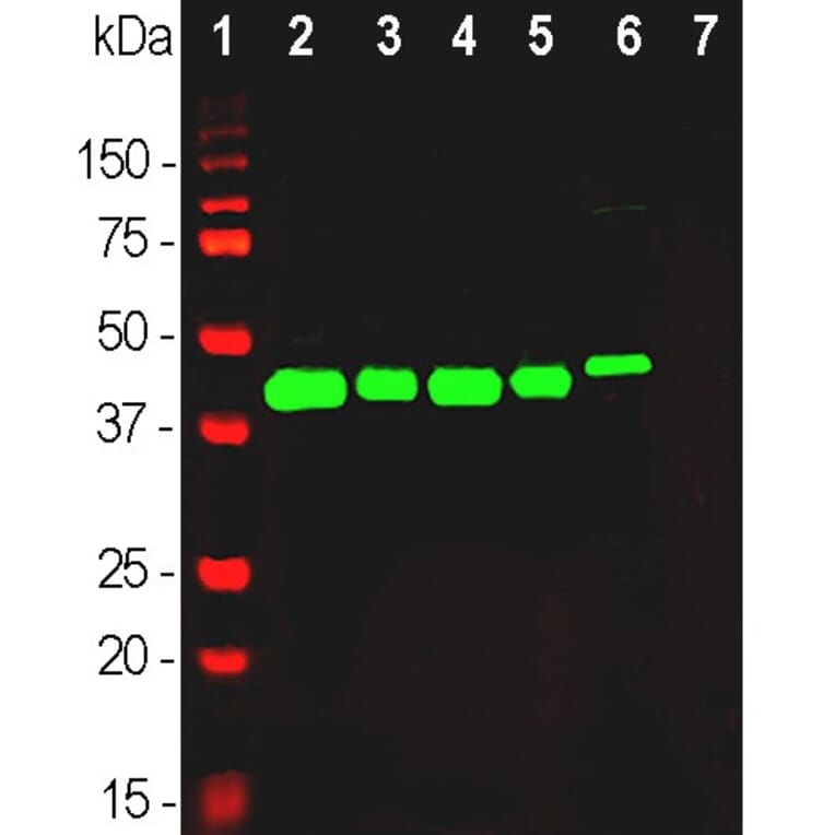
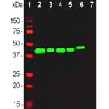
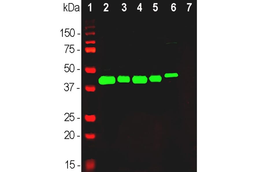
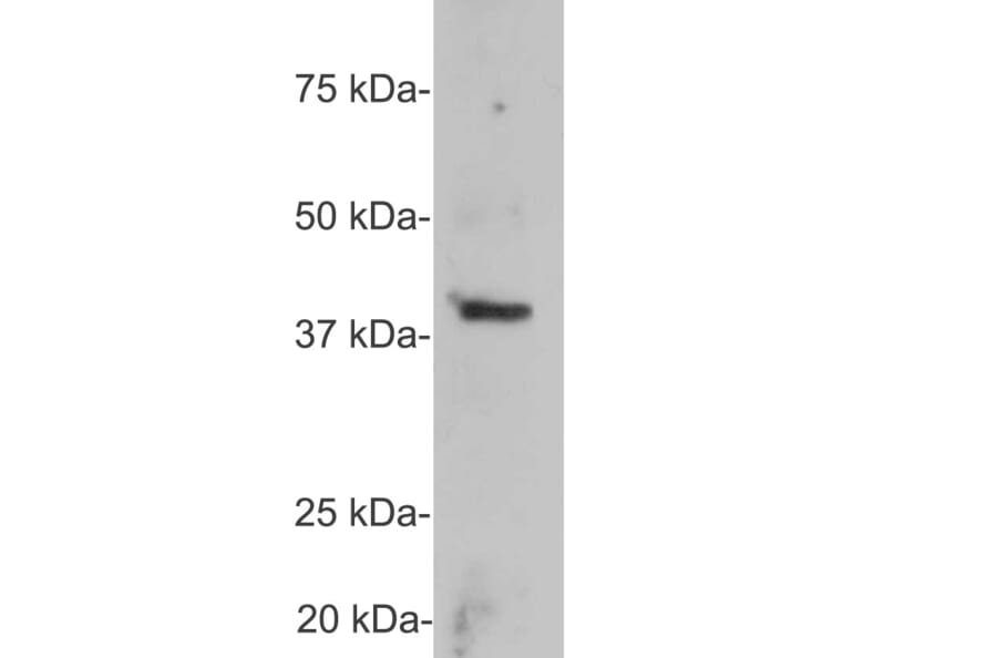
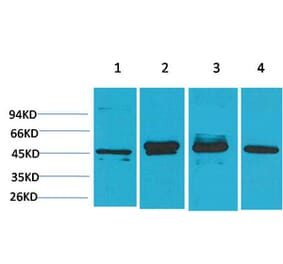
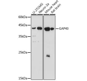
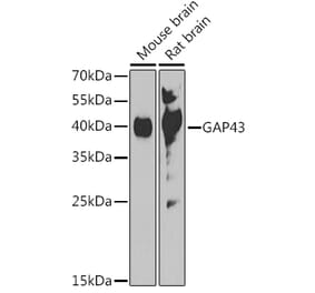
Western blot analysis of different tissue and cell lysates using Anti-GAP43 Antibody, at a dilution of 1:20,000, in green,: [Lane 1] protein standard (red), [Lane 2] rat brain, [Lane 3] rat spinal cord, [Lane 4] mouse brain, [Lane 5] mouse spinal cord, [Lane 6] SH-SY5Y cells, [Lane 7] C6 cells. Single band at 43 kDa mark corresponds to GAP43 protein. The GAP43 protein is detected only in the lysates of neuronal origin. C6 cells are a rat glioma cell line and do not express GAP43 protein.
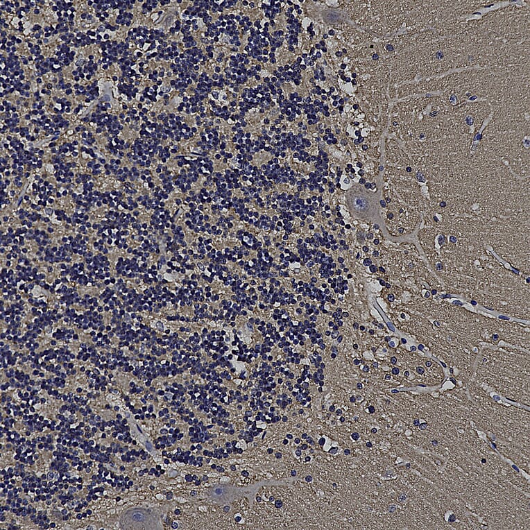
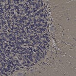
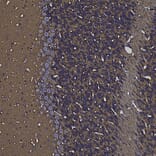
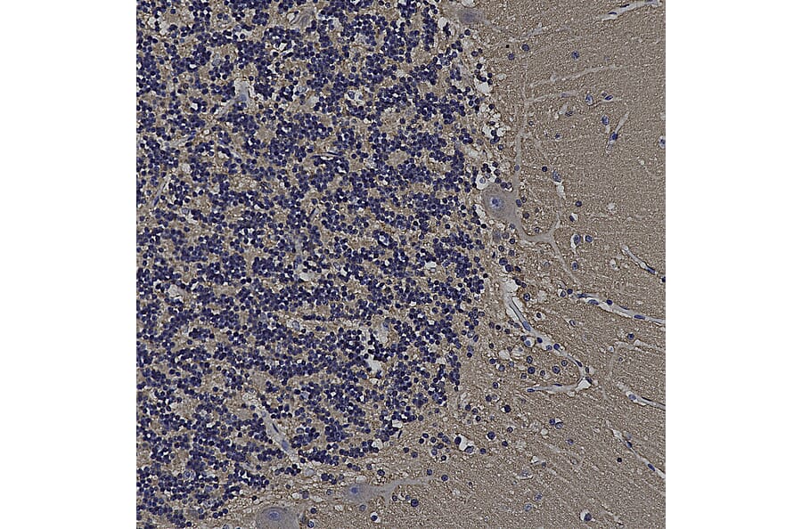
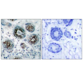
Immunohistochemistry analysis of a formalin fixed paraffin embedded human cerebellum section with Anti-GAP43 Antibody (A85394) at a dilution of 1:2,000 detected with DAB (brown) using the Vector Elite ABC-HRP detection and reagents with citra buffer retrieval. Counterstained with Hematoxylin (blue). The Anti-FABP7 Antibody (A104339) strongly labels cells and neuropil within the molecular layer and processes in the granular layer. Note: this antibody performs well in testing with both 4% PFA and standard NBF fixed rat, mouse and human tissues.
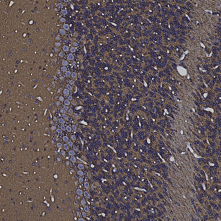
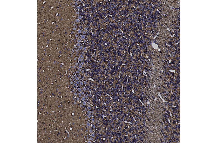
Immunohistochemistry analysis of a formalin fixed paraffin embedded mouse cerebellum section with Anti-GAP43 Antibody (A85394) at a dilution of 1:2,000 detected with DAB (brown) using the Vector Elite ABC-HRP detection and reagents with citra buffer retrieval. Counterstained with Hematoxylin (blue). The Anti-FABP7 Antibody (A104339) strongly labels cells and neuropil within the molecular layer and processes in the granular layer. Note: this antibody performs well in testing with both 4% PFA and standard NBF fixed rat, mouse and human tissues.


![Immunofluorescence - Anti-GAP43 Antibody [5E8] (A85392) - Antibodies.com](https://cdn.antibodies.com/image/catalog/85/A85392_1.jpg?profile=product_alternative)
![Immunofluorescence - Anti-GAP43 Antibody [3H14] (A85393) - Antibodies.com](https://cdn.antibodies.com/image/catalog/85/A85393_1.jpg?profile=product_alternative)
![Immunofluorescence - Anti-GAP43 Antibody [1E3] (A85391) - Antibodies.com](https://cdn.antibodies.com/image/catalog/85/A85391_1.jpg?profile=product_alternative)

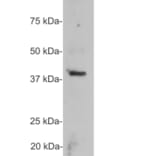
![Western Blot - Anti-GAP43 Antibody [ARC0246] (A305811) - Antibodies.com](https://cdn.antibodies.com/image/catalog/305/A305811_1.jpg?profile=product_alternative)