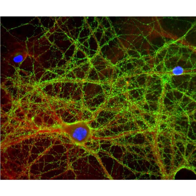
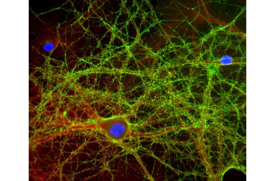
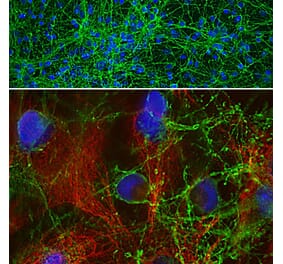
Mixed neuron-glial cultures stained with Anti-GAP43 Antibody (green) and Anti- rabbit antibody to alpha-II spectrin (A85352 | red), and DNA (blue). The Anti-GAP43 Antibody stains numerous axonal and dendritic profiles in these cultures, clearly revealing the submembraneous cytoskeleton and vesicles.
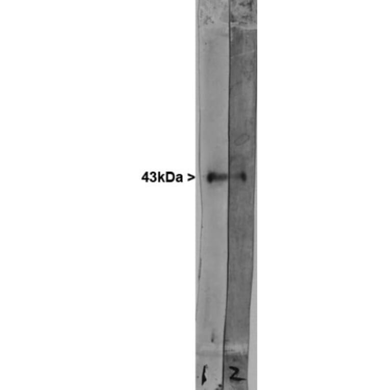
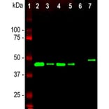
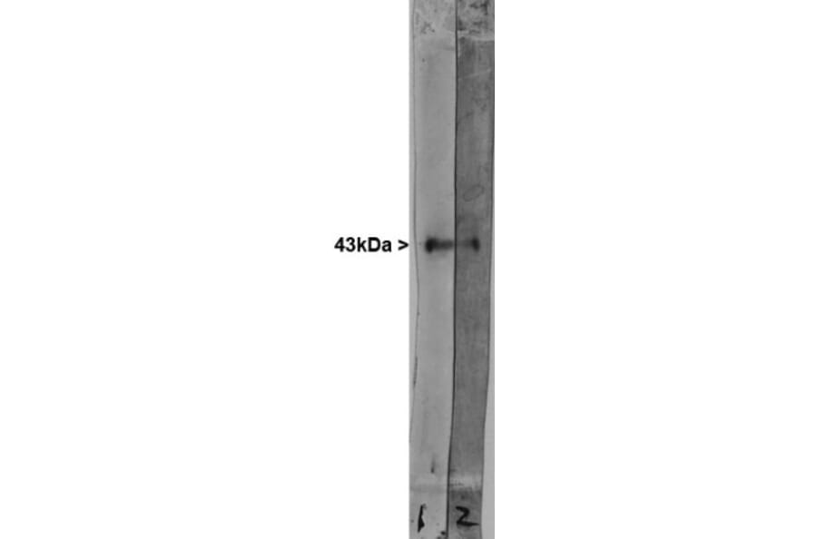
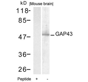
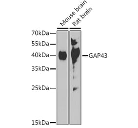
Western blot analysis of different tissue and cell lysates using Anti-GAP43 Antibody, at a dilution of 1:5,000, in green,: [Lane 1] protein standard (red), [Lane 2] rat brain, [Lane 3] rat spinal cord, [Lane 4] mouse brain, [Lane 5] mouse spinal cord, [Lane 6] C6 cells, [Lane 7] SH-SY5Y cells. Single band at the 43kDa mark corresponds to GAP43. The GAP43 protein only is detected in the lysates of neuronal origin. C6 cells are a rat glioma cell line and do not express GAP43.
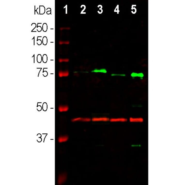
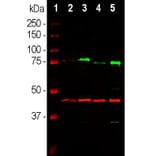
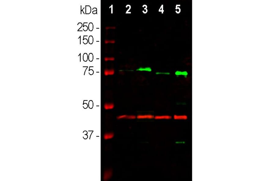
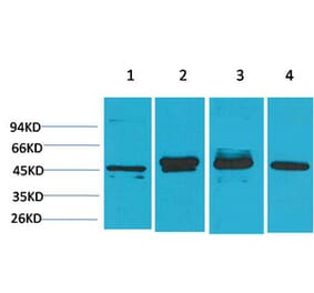
Western blot analysis of tissue lysates using Anti-MeCP2 Antibody [4F11] (A104324), at a dilution of 1:1,000, in green. The lanes contain: [Lane 1] protein standard (red), [Lane 2] rat whole brain, [Lane 3] nuclear fraction of rat brain, [Lane 4] mouse whole brain, and [Lane 5] nuclear fraction of mouse brain lysate. Strong band at about 75kDa in rat and slightly lower in mouse nuclear enriched fractions corresponds to the MeCP2 protein. The same blot was simultaneously probed with Anti-GAP43 Antibody (A85292) which detects GAP43 protein in all preparations with apparent molecular weight of 43kDa.
Publishing research using Anti-GAP43 Antibody (A85292)? Please let us know so that we can list the citation on this page.


![Immunofluorescence - Anti-GAP43 Antibody [5E8] (A85392) - Antibodies.com](https://cdn.antibodies.com/image/catalog/85/A85392_1.jpg?profile=product_alternative)
![Immunofluorescence - Anti-GAP43 Antibody [3H14] (A85393) - Antibodies.com](https://cdn.antibodies.com/image/catalog/85/A85393_1.jpg?profile=product_alternative)
![Immunofluorescence - Anti-GAP43 Antibody [1E3] (A85391) - Antibodies.com](https://cdn.antibodies.com/image/catalog/85/A85391_1.jpg?profile=product_alternative)

![Western Blot - Anti-GAP43 Antibody [ARC0246] (A305811) - Antibodies.com](https://cdn.antibodies.com/image/catalog/305/A305811_1.jpg?profile=product_alternative)