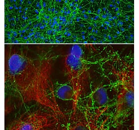
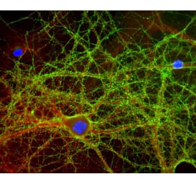
Immunofluorescent analysis of cortical neuron-glial cell culture from E20 rat stained with Anti-GAP43 Antibody, at a dilution of 1:1,000, in red, and Anti-MAP2 Antibody (A85363 | 1:10,000, in green. The blue is DAPI staining of nuclear DNA. Anti-GAP43 Antibody labels protein expressed in the axonal membrane of the neuronal cells, while the Anti-MAP2 Antibody stains dendrites and perikarya of neurons.
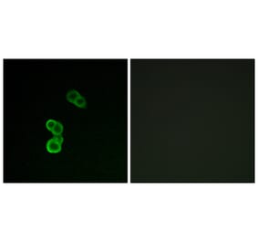
Mixed rat neuronal cultures stained with Anti-GAP43 Antibody (green). The Anti-GAP43 Antibody stains the plasma membrane of neurons and is particularly concentrated in dendrites.
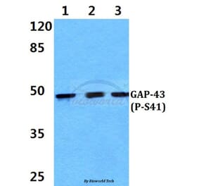
Western Blot - Anti-GAP43 Antibody [3H14] (A85393)
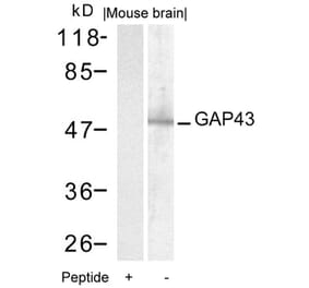
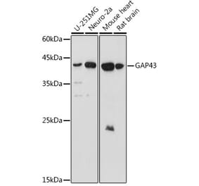
Western blot analysis of different tissue and cell lysates using Anti-GAP43 Antibody, at a dilution of 1:5,000, in green,: [Lane 1] protein standard (red), [Lane 2] rat brain, [Lane 3] rat spinal cord, [Lane 4] mouse brain, [Lane 5] mouse spinal cord, [Lane 6] C6 cells, [Lane 7] SH-SY5Y cells. The single band at the 43kDa mark corresponds to the GAP43 protein. The protein is expressed in rodent and human neurons and neuronal derived cells but not in C6 cells which are of glial origin.
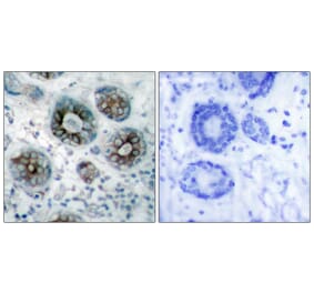
Immunohistochemistry analysis of a 4% PFA fixed paraffin embedded rat brain stem section with Anti-GAP43 Antibody [3H14] (A85393) at a dilution of 1:2,000 detected with DAB (brown) using the Vector Labs ImmPRESS method and reagents with citra buffer retrieval. Counterstained with Hematoxylin (blue). The Anti-GAP43 Antibody [3H14] (A85393) labels neurons (and to a lesser degree reactive glial cells) and is found concentrated in growth cones and axon terminals. Note: this antibody performs well in testing with 4% PFA or standard NBF fixed human and rat tissue.
Western Blot - Anti-GAP43 Antibody [3H14] (A85393)
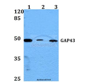
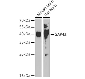
Western blot analysis of Anti-GAP43 Antibody. Blot of SH-SY5Y cell lysate (Lane 1) and whole rat brain lysate (Lane 2) was probed with Anti-GAP43 Antibody, at a dilution of 1: 2,000). Note, the strong single band running at about 43kDa corresponding to GAP43.


![Immunofluorescence - Anti-GAP43 Antibody [3H14] (A85393) - Antibodies.com](https://cdn.antibodies.com/image/catalog/85/A85393_1.jpg?profile=product_top)
![Immunofluorescence - Anti-GAP43 Antibody [3H14] (A85393) - Antibodies.com](https://cdn.antibodies.com/image/catalog/85/A85393_2.jpg?profile=product_top)
![Western Blot - Anti-GAP43 Antibody [3H14] (A85393) - Antibodies.com](https://cdn.antibodies.com/image/catalog/85/A85393_3.jpg?profile=product_top)
![Immunohistochemistry - Anti-GAP43 Antibody [3H14] (A85393) - Antibodies.com](https://cdn.antibodies.com/image/catalog/85/A85393_4.jpg?profile=product_top)
![Western Blot - Anti-GAP43 Antibody [3H14] (A85393) - Antibodies.com](https://cdn.antibodies.com/image/catalog/85/A85393_5.jpg?profile=product_top)
![Immunofluorescence - Anti-GAP43 Antibody [3H14] (A85393) - Antibodies.com](https://cdn.antibodies.com/image/catalog/85/A85393_1.jpg?profile=product_top_thumb)
![Immunofluorescence - Anti-GAP43 Antibody [3H14] (A85393) - Antibodies.com](https://cdn.antibodies.com/image/catalog/85/A85393_2.jpg?profile=product_top_thumb)
![Western Blot - Anti-GAP43 Antibody [3H14] (A85393) - Antibodies.com](https://cdn.antibodies.com/image/catalog/85/A85393_3.jpg?profile=product_top_thumb)
![Immunohistochemistry - Anti-GAP43 Antibody [3H14] (A85393) - Antibodies.com](https://cdn.antibodies.com/image/catalog/85/A85393_4.jpg?profile=product_top_thumb)
![Western Blot - Anti-GAP43 Antibody [3H14] (A85393) - Antibodies.com](https://cdn.antibodies.com/image/catalog/85/A85393_5.jpg?profile=product_top_thumb)
![Immunofluorescence - Anti-GAP43 Antibody [3H14] (A85393) - Antibodies.com](https://cdn.antibodies.com/image/catalog/85/A85393_1.jpg?profile=product_image)
![Immunofluorescence - Anti-GAP43 Antibody [3H14] (A85393) - Antibodies.com](https://cdn.antibodies.com/image/catalog/85/A85393_2.jpg?profile=product_image)
![Western Blot - Anti-GAP43 Antibody [3H14] (A85393) - Antibodies.com](https://cdn.antibodies.com/image/catalog/85/A85393_3.jpg?profile=product_image)
![Immunohistochemistry - Anti-GAP43 Antibody [3H14] (A85393) - Antibodies.com](https://cdn.antibodies.com/image/catalog/85/A85393_4.jpg?profile=product_image)
![Western Blot - Anti-GAP43 Antibody [3H14] (A85393) - Antibodies.com](https://cdn.antibodies.com/image/catalog/85/A85393_5.jpg?profile=product_image)
![Immunofluorescence - Anti-GAP43 Antibody [5E8] (A85392) - Antibodies.com](https://cdn.antibodies.com/image/catalog/85/A85392_1.jpg?profile=product_alternative)
![Immunofluorescence - Anti-GAP43 Antibody [1E3] (A85391) - Antibodies.com](https://cdn.antibodies.com/image/catalog/85/A85391_1.jpg?profile=product_alternative)

![Western Blot - Anti-GAP43 Antibody [ARC0246] (A305811) - Antibodies.com](https://cdn.antibodies.com/image/catalog/305/A305811_1.jpg?profile=product_alternative)