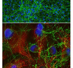
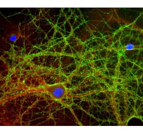
Immunofluorescent analysis of mixed neuronal cultures, co-stained with Anti-GAP43 Antibody [1E3] (A85391) (green), a rabbit antibody to microtubule associated protein 2 (MAP2) (red), and a stain for DNA (blue). The GAP43 antibody stains the plasma membrane of neurons and is particularly concentrated in dendrites.
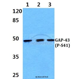
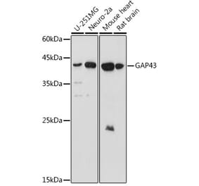
Western blot of whole rat spinal cord lysates probed with Anti-GAP43 Antibody [1E3] (A85391) to GAP43 in lane 16 and another similar antibody in lane 17. Dots in middle of strips indicate position of 50kDa and 37kDa protein bands. The strong single band running at about 43kDa corresponds to GAP43.
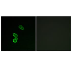
Mixed neuronal cultures stained with Anti-GAP43 Antibody (green), Anti-MAP2 Antibody (A85363 | red) and DNA (blue). The Anti-GAP43 Antibody stains the plasma membrane of neurons and is particularly concentrated in dendrites.
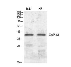
Western blot of whole rat spinal cord lysates probed with Anti-GAP43 Antibody in Lane 16 and another similar antibody in Lane 17. Dots in middle of strips indicate position of 50kDa and 37kDa protein bands. Note that the strong single band running at about 43kDa corresponds to GAP43.
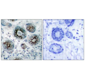
Immunohistochemistry analysis of a NBF fixed paraffin embedded human cerebellum section with Anti-GAP43 Antibody [1E3] (A85391) at a dilution of 1:2,000 detected in DAB (brown) using Vector Labs ImmPRESS method and reagents with citra buffer retrieval. Counterstained with Hematoxylin (blue). Anti-GAP43 Antibody [1E3] (A85391) labels molecular layer cells and neuropil as well as processes in the granular layer. Note: this antibody performs well in testing with both 4% PFA and standard NBF fixed tissues.


![Immunofluorescence - Anti-GAP43 Antibody [1E3] (A85391) - Antibodies.com](https://cdn.antibodies.com/image/catalog/85/A85391_1.jpg?profile=product_top)
![Western Blot - Anti-GAP43 Antibody [1E3] (A85391) - Antibodies.com](https://cdn.antibodies.com/image/catalog/85/A85391_2.jpg?profile=product_top)
![Immunofluorescence - Anti-GAP43 Antibody [1E3] (A85391) - Antibodies.com](https://cdn.antibodies.com/image/catalog/85/A85391_3.jpg?profile=product_top)
![Western Blot - Anti-GAP43 Antibody [1E3] (A85391) - Antibodies.com](https://cdn.antibodies.com/image/catalog/85/A85391_4.jpg?profile=product_top)
![Immunohistochemistry - Anti-GAP43 Antibody [1E3] (A85391) - Antibodies.com](https://cdn.antibodies.com/image/catalog/85/A85391_5.jpg?profile=product_top)
![Immunofluorescence - Anti-GAP43 Antibody [1E3] (A85391) - Antibodies.com](https://cdn.antibodies.com/image/catalog/85/A85391_1.jpg?profile=product_top_thumb)
![Western Blot - Anti-GAP43 Antibody [1E3] (A85391) - Antibodies.com](https://cdn.antibodies.com/image/catalog/85/A85391_2.jpg?profile=product_top_thumb)
![Immunofluorescence - Anti-GAP43 Antibody [1E3] (A85391) - Antibodies.com](https://cdn.antibodies.com/image/catalog/85/A85391_3.jpg?profile=product_top_thumb)
![Western Blot - Anti-GAP43 Antibody [1E3] (A85391) - Antibodies.com](https://cdn.antibodies.com/image/catalog/85/A85391_4.jpg?profile=product_top_thumb)
![Immunohistochemistry - Anti-GAP43 Antibody [1E3] (A85391) - Antibodies.com](https://cdn.antibodies.com/image/catalog/85/A85391_5.jpg?profile=product_top_thumb)
![Immunofluorescence - Anti-GAP43 Antibody [1E3] (A85391) - Antibodies.com](https://cdn.antibodies.com/image/catalog/85/A85391_1.jpg?profile=product_image)
![Western Blot - Anti-GAP43 Antibody [1E3] (A85391) - Antibodies.com](https://cdn.antibodies.com/image/catalog/85/A85391_2.jpg?profile=product_image)
![Immunofluorescence - Anti-GAP43 Antibody [1E3] (A85391) - Antibodies.com](https://cdn.antibodies.com/image/catalog/85/A85391_3.jpg?profile=product_image)
![Western Blot - Anti-GAP43 Antibody [1E3] (A85391) - Antibodies.com](https://cdn.antibodies.com/image/catalog/85/A85391_4.jpg?profile=product_image)
![Immunohistochemistry - Anti-GAP43 Antibody [1E3] (A85391) - Antibodies.com](https://cdn.antibodies.com/image/catalog/85/A85391_5.jpg?profile=product_image)
![Immunofluorescence - Anti-GAP43 Antibody [5E8] (A85392) - Antibodies.com](https://cdn.antibodies.com/image/catalog/85/A85392_1.jpg?profile=product_alternative)
![Immunofluorescence - Anti-GAP43 Antibody [3H14] (A85393) - Antibodies.com](https://cdn.antibodies.com/image/catalog/85/A85393_1.jpg?profile=product_alternative)

![Western Blot - Anti-GAP43 Antibody [ARC0246] (A305811) - Antibodies.com](https://cdn.antibodies.com/image/catalog/305/A305811_1.jpg?profile=product_alternative)