+1 (314) 370-6046 or
Contact Us - Argentina
- Australia
- Austria
- Bahrain
- Belgium
- Brazil
- Bulgaria
- Cameroon
- Canada
- Chile
- China
- Colombia
- Croatia
- Cyprus
- Czech Republic
- Denmark
- Ecuador
- Egypt
- Estonia
- Finland
- France
- Germany
- Greece
- Hong Kong
- Hungary
- Iceland
- India
- Indonesia
- Iran
- Ireland
- Israel
- Italy
- Japan
- Kazakhstan
- Kuwait
- Latvia
- Lithuania
- Luxembourg
- Macedonia
- Malaysia
- Malta
- Mexico
- Monaco
- Morocco
- Netherlands
- New Zealand
- Nigeria
- Norway
- Peru
- Philippines
- Poland
- Portugal
- Qatar
- Romania
- Russia
- Saudi Arabia
- Serbia
- Singapore
- Slovakia
- Slovenia
- South Africa
- South Korea
- Spain
- Sri Lanka
- Sweden
- Switzerland
- Taiwan
- Thailand
- Turkey
- Ukraine
- UAE
- United Kingdom
- United States
- Venezuela
- Vietnam

![Immunohistochemistry - Anti-Galectin 3 Antibody [5C21] (A85368) - Antibodies.com](https://cdn.antibodies.com/image/catalog/85/A85368_1.jpg?profile=product_top)
![Immunofluorescence - Anti-Galectin 3 Antibody [5C21] (A85368) - Antibodies.com](https://cdn.antibodies.com/image/catalog/85/A85368_2.jpg?profile=product_top)
![Immunofluorescence - Anti-Galectin 3 Antibody [5C21] (A85368) - Antibodies.com](https://cdn.antibodies.com/image/catalog/85/A85368_3.jpg?profile=product_top)
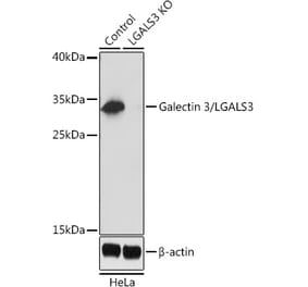
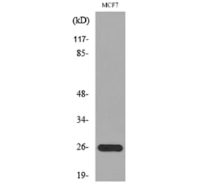
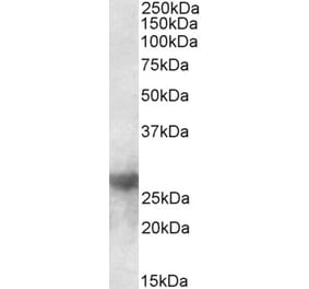
![Western Blot - Anti-Galectin 3 Antibody [5C21] (A85368) - Antibodies.com](https://cdn.antibodies.com/image/catalog/85/A85368_4.jpg?profile=product_top)
![Western Blot - Anti-Galectin 3 Antibody [5C21] (A85368) - Antibodies.com](https://cdn.antibodies.com/image/catalog/85/A85368_5.jpg?profile=product_top)
![Immunohistochemistry - Anti-Galectin 3 Antibody [5C21] (A85368) - Antibodies.com](https://cdn.antibodies.com/image/catalog/85/A85368_1.jpg?profile=product_top_thumb)
![Immunofluorescence - Anti-Galectin 3 Antibody [5C21] (A85368) - Antibodies.com](https://cdn.antibodies.com/image/catalog/85/A85368_2.jpg?profile=product_top_thumb)
![Immunofluorescence - Anti-Galectin 3 Antibody [5C21] (A85368) - Antibodies.com](https://cdn.antibodies.com/image/catalog/85/A85368_3.jpg?profile=product_top_thumb)
![Western Blot - Anti-Galectin 3 Antibody [5C21] (A85368) - Antibodies.com](https://cdn.antibodies.com/image/catalog/85/A85368_4.jpg?profile=product_top_thumb)
![Western Blot - Anti-Galectin 3 Antibody [5C21] (A85368) - Antibodies.com](https://cdn.antibodies.com/image/catalog/85/A85368_5.jpg?profile=product_top_thumb)
![Immunohistochemistry - Anti-Galectin 3 Antibody [5C21] (A85368) - Antibodies.com](https://cdn.antibodies.com/image/catalog/85/A85368_1.jpg?profile=product_image)
![Immunofluorescence - Anti-Galectin 3 Antibody [5C21] (A85368) - Antibodies.com](https://cdn.antibodies.com/image/catalog/85/A85368_2.jpg?profile=product_image)
![Immunofluorescence - Anti-Galectin 3 Antibody [5C21] (A85368) - Antibodies.com](https://cdn.antibodies.com/image/catalog/85/A85368_3.jpg?profile=product_image)
![Western Blot - Anti-Galectin 3 Antibody [5C21] (A85368) - Antibodies.com](https://cdn.antibodies.com/image/catalog/85/A85368_4.jpg?profile=product_image)
![Western Blot - Anti-Galectin 3 Antibody [5C21] (A85368) - Antibodies.com](https://cdn.antibodies.com/image/catalog/85/A85368_5.jpg?profile=product_image)
![Immunohistochemistry - Anti-Galectin 3 Antibody [LGALS3/4792] (A277691) - Antibodies.com](https://cdn.antibodies.com/image/catalog/277/A277691_1.jpg?profile=product_alternative)
![Immunohistochemistry - Anti-Galectin 3 Antibody [LGALS3/4792] - BSA and Azide free (A278279) - Antibodies.com](https://cdn.antibodies.com/image/catalog/278/A278279_1.jpg?profile=product_alternative)

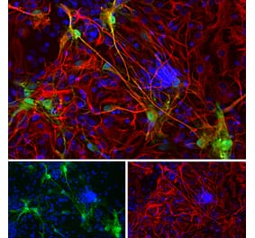
![Flow Cytometry - Anti-Galectin 3 Antibody [M3/38] (A285836) - Antibodies.com](https://cdn.antibodies.com/image/catalog/285/A285837_1.jpg?profile=product_alternative)
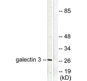
![Western Blot - Anti-Galectin 3 Antibody [ARC0542] (A306857) - Antibodies.com](https://cdn.antibodies.com/image/catalog/306/A306857_1.jpg?profile=product_alternative)
