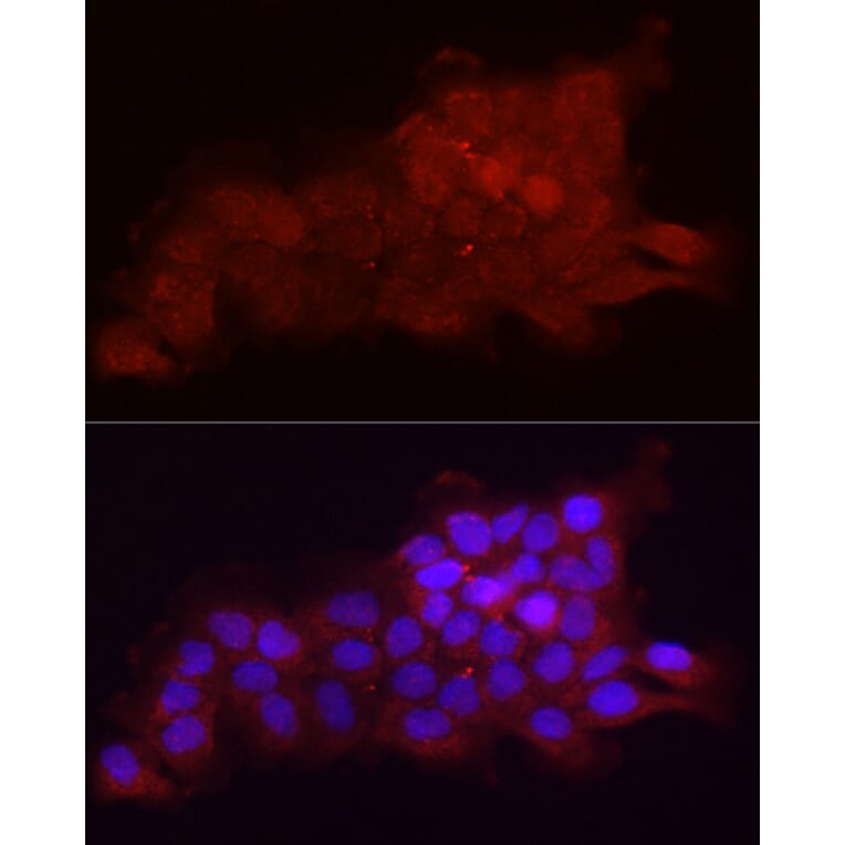
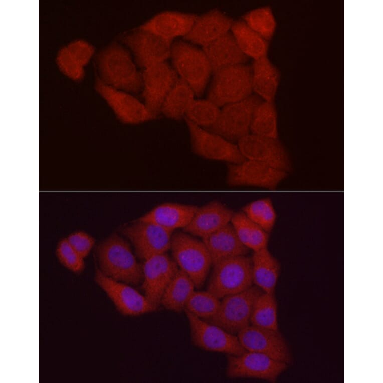
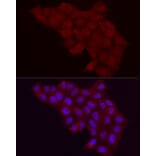
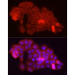
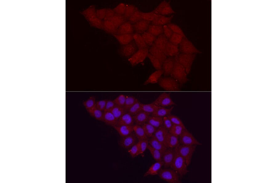
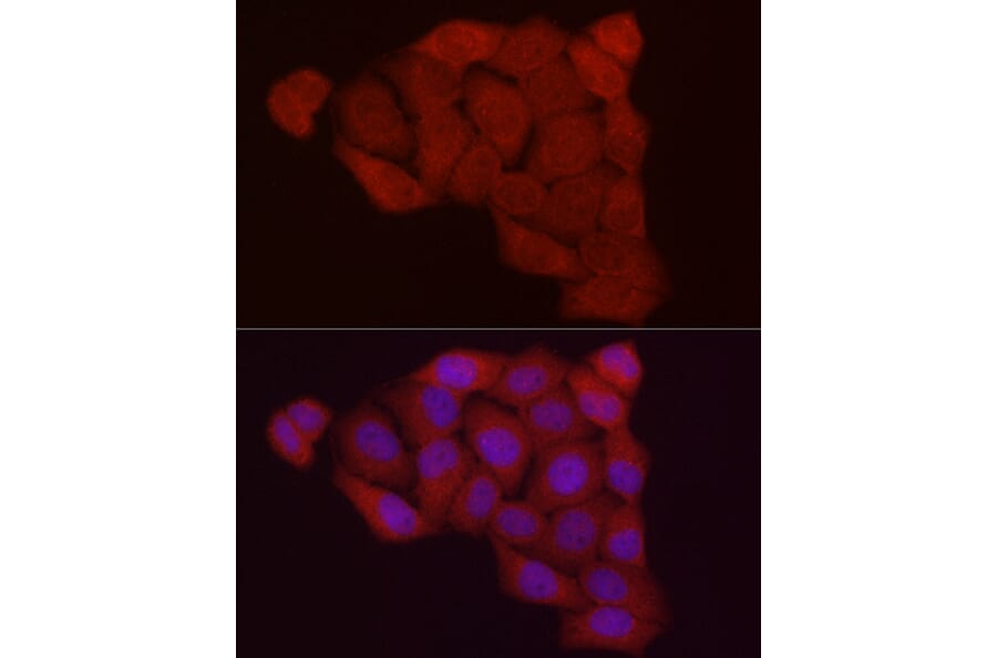
Fbx32 expression in A431 cells analyzed by immunofluorescence. Staining was performed with Anti-Fbx32 Antibody (A80674) at a dilution of 1:20 followed by Cy3 Goat Anti-Rabbit IgG (H+L) secondary antibody at a dilution of 1:500. Nuclei were stained with DAPI (blue).
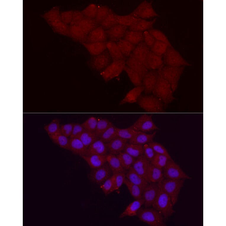
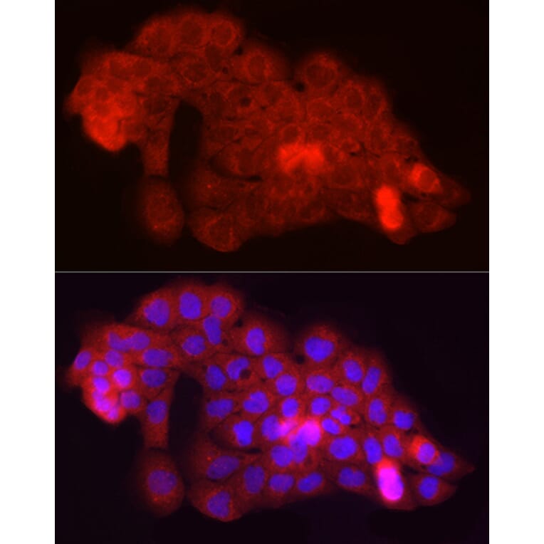
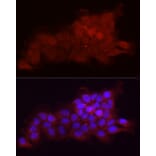
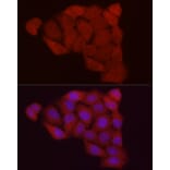
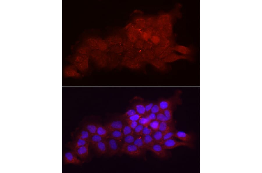
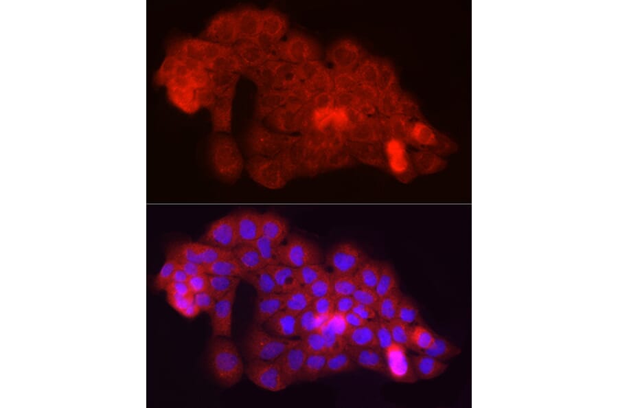
Fbx32 expression in Hela cells analyzed by immunofluorescence. Staining was performed with Anti-Fbx32 Antibody (A80674) at a dilution of 1:20 followed by Cy3 Goat Anti-Rabbit IgG (H+L) secondary antibody at a dilution of 1:500. Nuclei were stained with DAPI (blue).
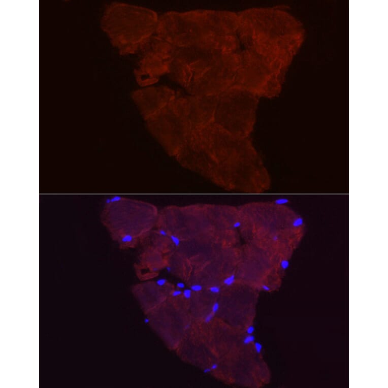
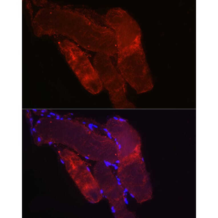
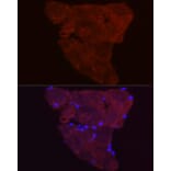
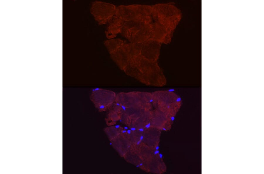
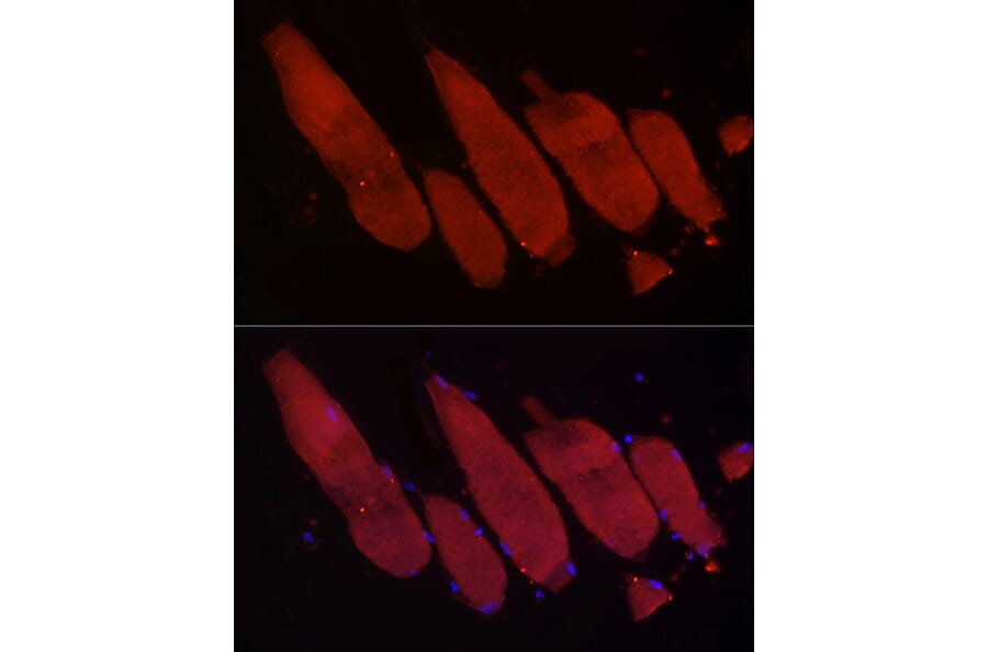
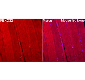
Fbx32 expression in paraffin-embedded Mouse skeletal muscle tissue analyzed by immunofluorescence. Staining was performed with Anti-Fbx32 Antibody (A80674) at a dilution of 1:20 followed by Cy3 Goat Anti-Rabbit IgG (H+L) secondary antibody at a dilution of 1:500. Nuclei were stained with DAPI (blue).
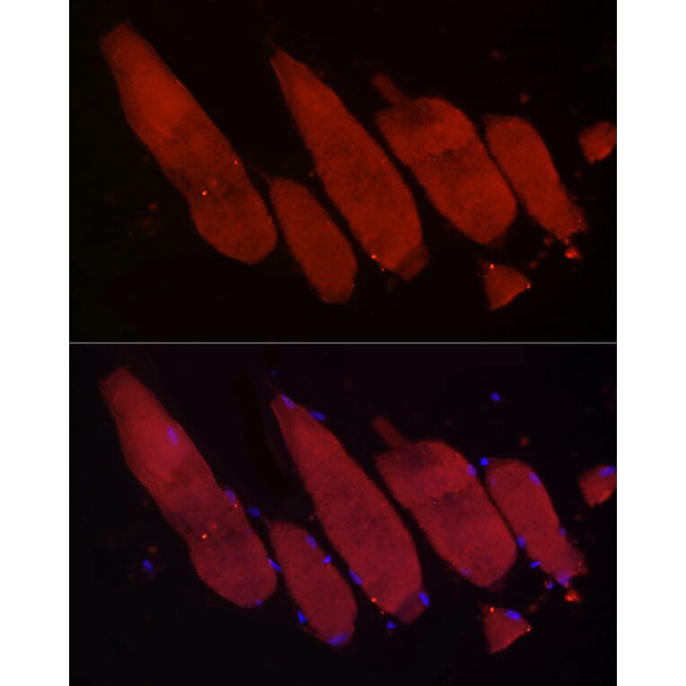
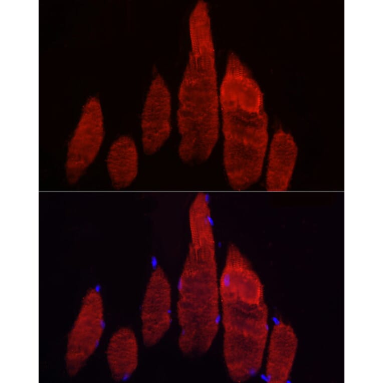
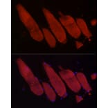
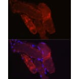
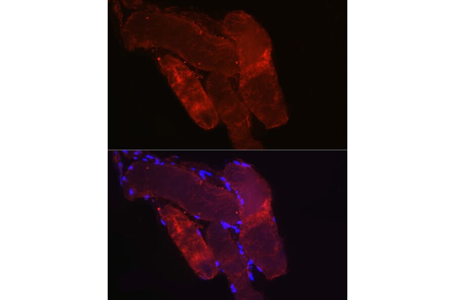
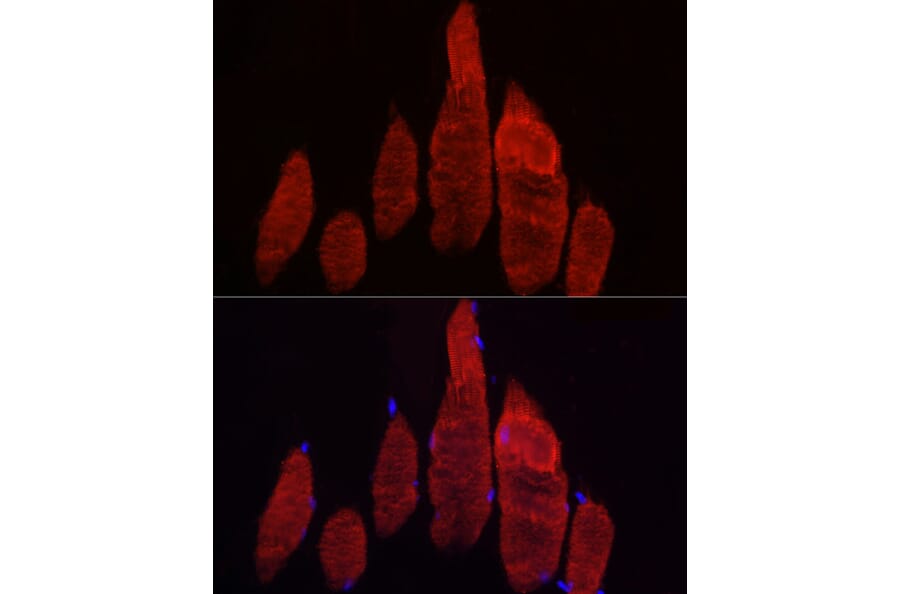
Fbx32 expression in paraffin-embedded Rat skeletal muscle tissue analyzed by immunofluorescence. Staining was performed with Anti-Fbx32 Antibody (A80674) at a dilution of 1:20 followed by Cy3 Goat Anti-Rabbit IgG (H+L) secondary antibody at a dilution of 1:500. Nuclei were stained with DAPI (blue).
Fbx32 expression in A431 cells analyzed by immunofluorescence. Staining was performed with Anti-Fbx32 Antibody (A80674) at a dilution of 1:20 followed by Cy3 Goat Anti-Rabbit IgG (H+L) secondary antibody at a dilution of 1:500. Nuclei were stained with DAPI (blue).
Fbx32 expression in Hela cells analyzed by immunofluorescence. Staining was performed with Anti-Fbx32 Antibody (A80674) at a dilution of 1:20 followed by Cy3 Goat Anti-Rabbit IgG (H+L) secondary antibody at a dilution of 1:500. Nuclei were stained with DAPI (blue).
Fbx32 expression in paraffin-embedded Mouse skeletal muscle tissue analyzed by immunofluorescence. Staining was performed with Anti-Fbx32 Antibody (A80674) at a dilution of 1:20 followed by Cy3 Goat Anti-Rabbit IgG (H+L) secondary antibody at a dilution of 1:500. Nuclei were stained with DAPI (blue).
Fbx32 expression in paraffin-embedded Rat skeletal muscle tissue analyzed by immunofluorescence. Staining was performed with Anti-Fbx32 Antibody (A80674) at a dilution of 1:20 followed by Cy3 Goat Anti-Rabbit IgG (H+L) secondary antibody at a dilution of 1:500. Nuclei were stained with DAPI (blue).


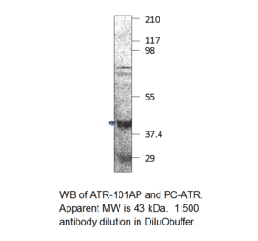
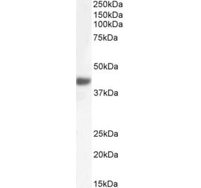
![Western Blot - Anti-Fbx32 Antibody [ARC0830] (A307603) - Antibodies.com](https://cdn.antibodies.com/image/catalog/307/A307603_1.jpg?profile=product_alternative)