Supplied in Phosphate Buffered Saline, pH 7.3, with 50% Glycerol and 0.02% Sodium Azide.
Storage
Shipped at 4°C. Upon delivery aliquot and store at -20°C. Avoid freeze/thaw cycles.
Synonyms
Damage-specific DNA-binding protein 1, DDB p127 subunit, DDBa, DNA damage-binding protein 1, DNA damage-binding protein a, HBV X-associated protein 1, UV-damaged DNA-binding factor, XAP-1, XAP1
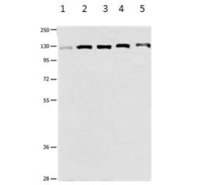
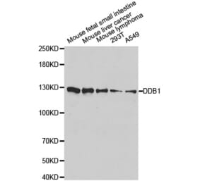
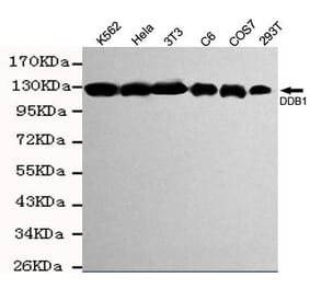
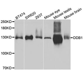
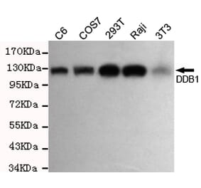
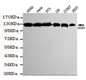
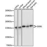
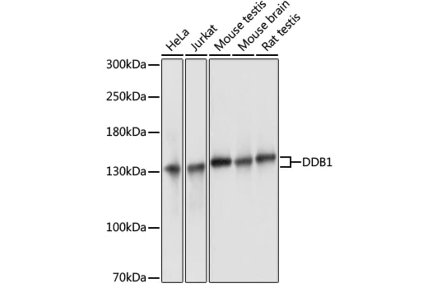
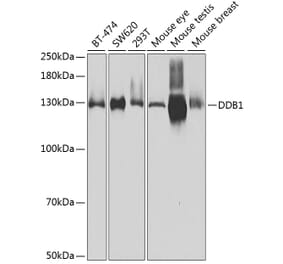
Western blot analysis of extracts of various cell lines, using Anti-DDB1 Antibody (A88091) at 1:3,000 dilution. The secondary antibody was Goat Anti-Rabbit IgG H&L Antibody (HRP) at 1:10,000 dilution. Lysates/proteins were present at 25µg per lane. The blocking buffer used was 3% non-fat dry milk in TBST. Detection was with a ECL Basic Kit. Exposure time: 1s.
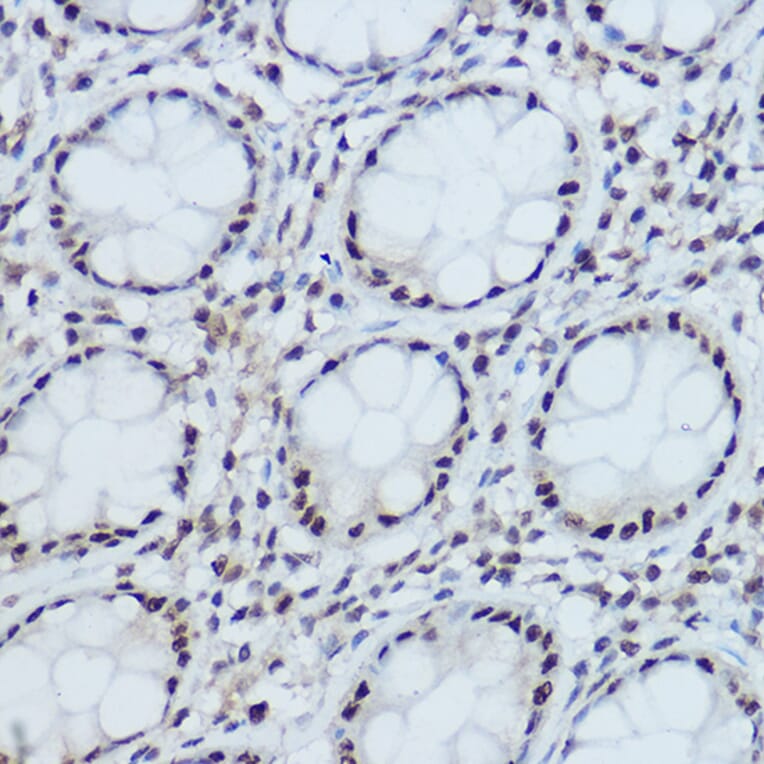
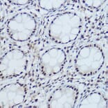
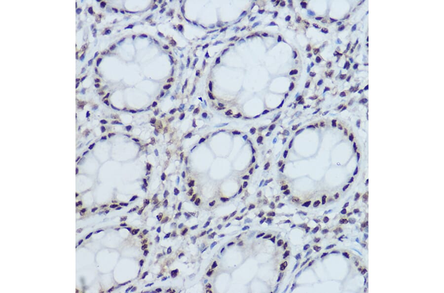
Immunohistochemistry analysis of paraffin-embedded human colon tissue using Anti-DDB1 Antibody (A88091) at a dilution of 1:300 (40x lens). Perform high pressure antigen retrieval with 10 mM citrate buffer pH 6.0 before commencing with IHC staining protocol.
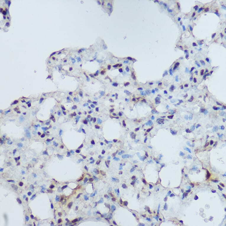
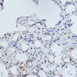
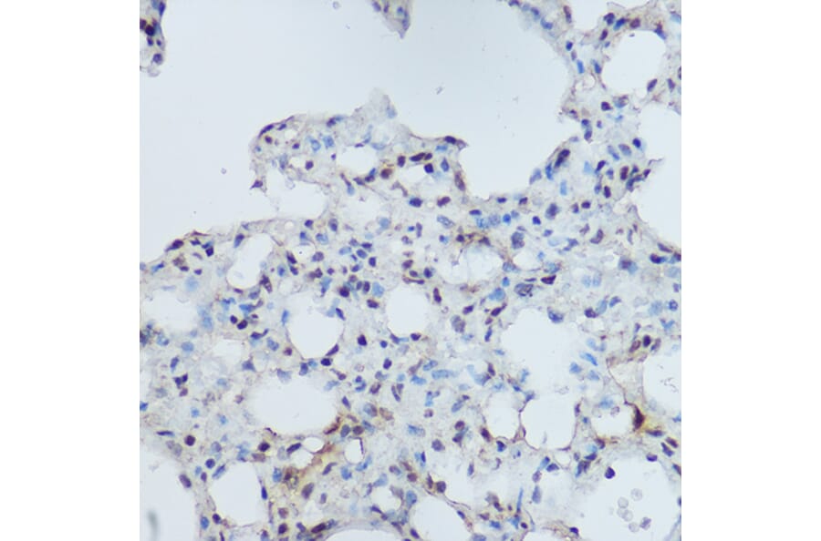
Immunohistochemistry analysis of paraffin-embedded mouse lung using Anti-DDB1 Antibody (A88091) at a dilution of 1:300 (40x lens). Perform high pressure antigen retrieval with 10 mM citrate buffer pH 6.0 before commencing with IHC staining protocol.
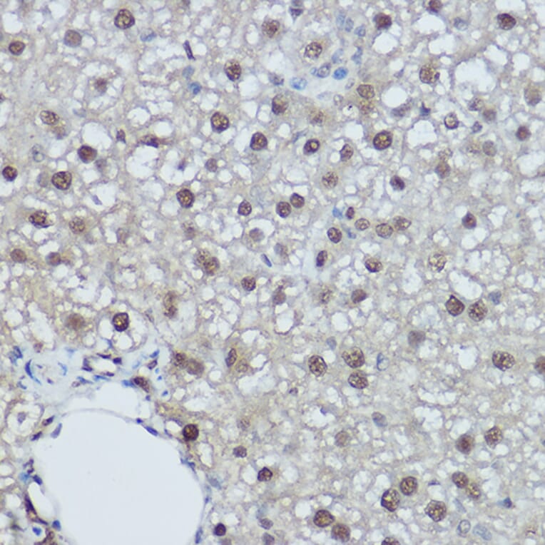
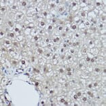
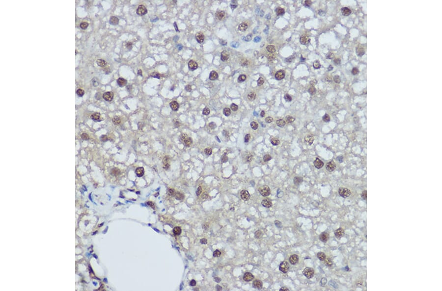
Immunohistochemistry analysis of paraffin-embedded rat liver using Anti-DDB1 Antibody (A88091) at a dilution of 1:300 (40x lens). Perform high pressure antigen retrieval with 10 mM citrate buffer pH 6.0 before commencing with IHC staining protocol.
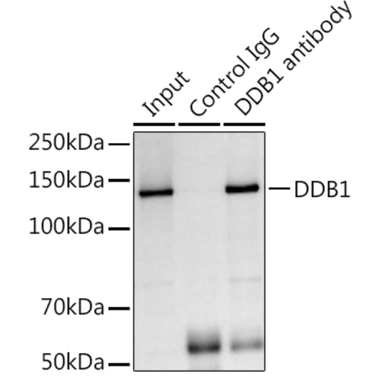
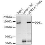
Immunoprecipitation analysis of 300µg extracts of Jurkat cells using 3µg of Anti-DDB1 Antibody (A88091). This Western blot was performed on the immunoprecipitate using Anti-DDB1 Antibody (A88091) at a dilution of 1:1000.


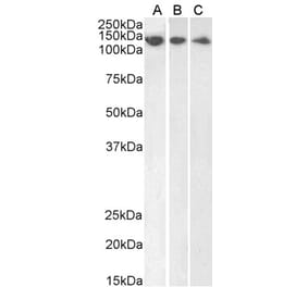
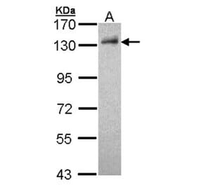
![Western Blot - Anti-DDB1 Antibody [ARC1278] (A308971) - Antibodies.com](https://cdn.antibodies.com/image/catalog/308/A308971_1.jpg?profile=product_alternative)