Supplied in Phosphate Buffered Saline, pH 7.3, with 50% Glycerol and 0.01% Thiomersal.
Storage
Shipped at 4°C. Upon delivery aliquot and store at -20°C. Avoid freeze/thaw cycles.
Synonyms
Damage-specific DNA-binding protein 1, DDB p127 subunit, DDBa, DNA damage-binding protein 1, DNA damage-binding protein a, HBV X-associated protein 1, UV-damaged DNA-binding factor, XAP-1, XAP1
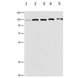
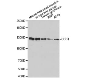
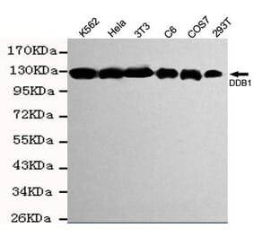
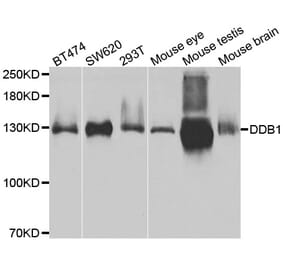
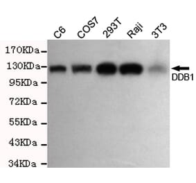
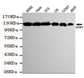
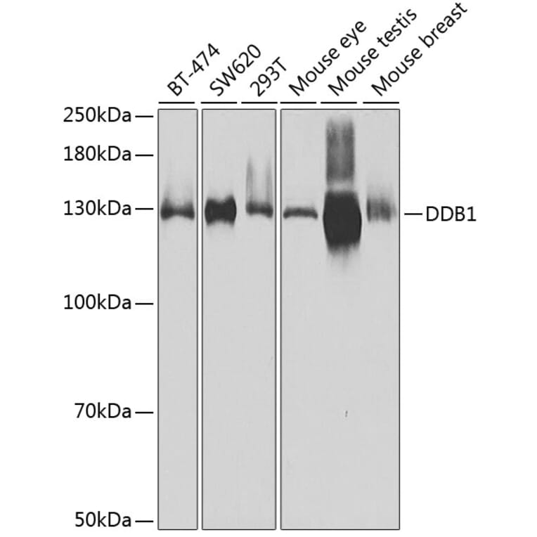
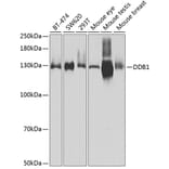
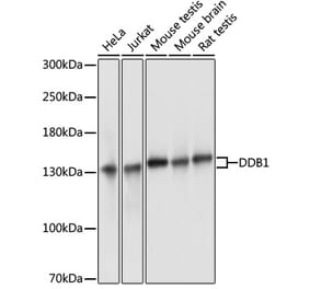
Western blot analysis of extracts of various cell lines, using Anti-DDB1 Antibody (A14242) at 1:500 dilution. The secondary antibody was Goat Anti-Rabbit IgG H&L Antibody (HRP) at 1:10,000 dilution. Lysates/proteins were present at 25µg per lane. The blocking buffer used was 3% non-fat dry milk in TBST. Detection was with a ECL Basic Kit. Exposure time: 30s.
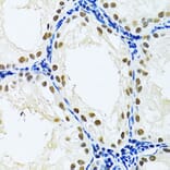
Immunohistochemistry analysis of paraffin-embedded rat testis using Anti-DDB1 Antibody (A14242) at a dilution of 1:100 (40x lens). Perform microwave antigen retrieval with 10 mM PBS buffer pH 7.2 before commencing with IHC staining protocol.
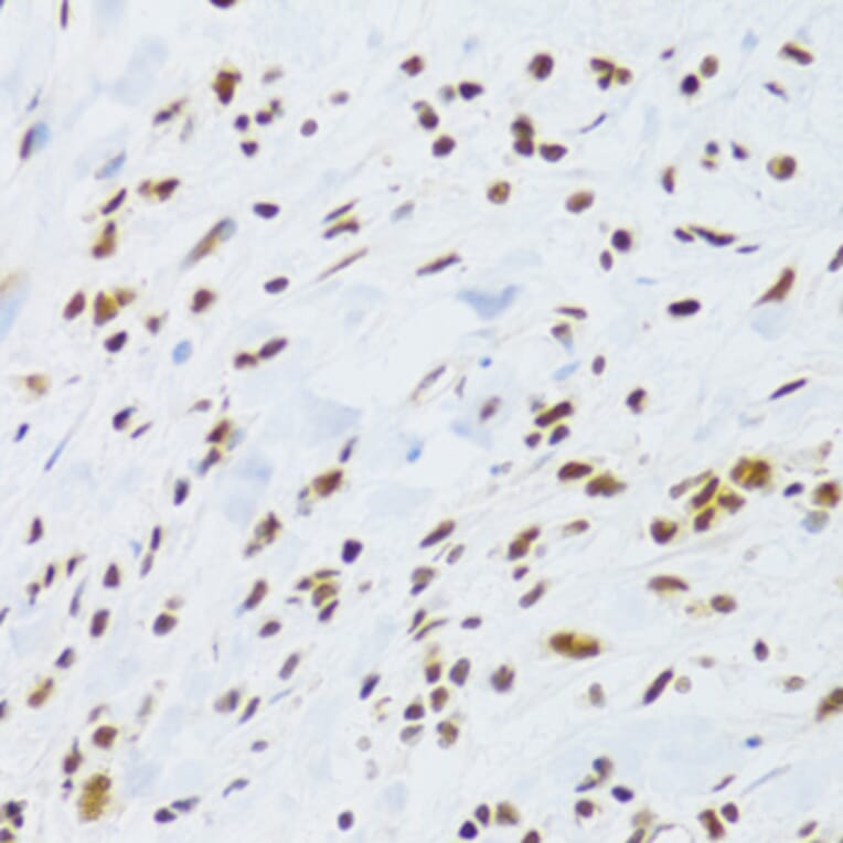
Immunohistochemistry analysis of paraffin-embedded human gastric cancer using Anti-DDB1 Antibody (A14242) at a dilution of 1:100 (40x lens). Perform microwave antigen retrieval with 10 mM PBS buffer pH 7.2 before commencing with IHC staining protocol.
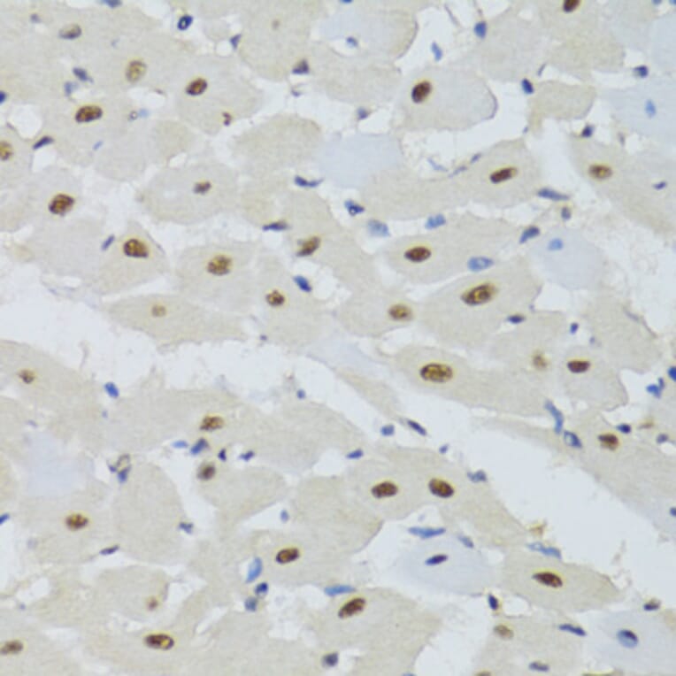
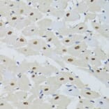
Immunohistochemistry analysis of paraffin-embedded mouse heart using Anti-DDB1 Antibody (A14242) at a dilution of 1:100 (40x lens). Perform microwave antigen retrieval with 10 mM PBS buffer pH 7.2 before commencing with IHC staining protocol.
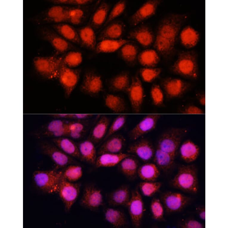
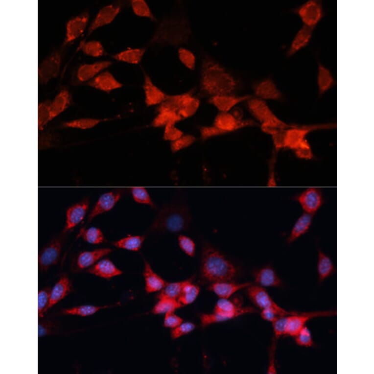
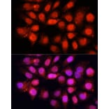
Immunofluorescence analysis of HeLa cells using Anti-DDB1 Antibody (A14242) at a dilution of 1:100 (40x lens). DAPI was used to stain the cell nuclei (blue).
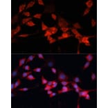
Immunofluorescence analysis of NIH-3T3 cells using Anti-DDB1 Antibody (A14242) at a dilution of 1:100 (40x lens). DAPI was used to stain the cell nuclei (blue).


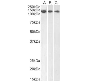
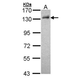
![Western Blot - Anti-DDB1 Antibody [ARC1278] (A308971) - Antibodies.com](https://cdn.antibodies.com/image/catalog/308/A308971_1.jpg?profile=product_alternative)