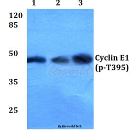
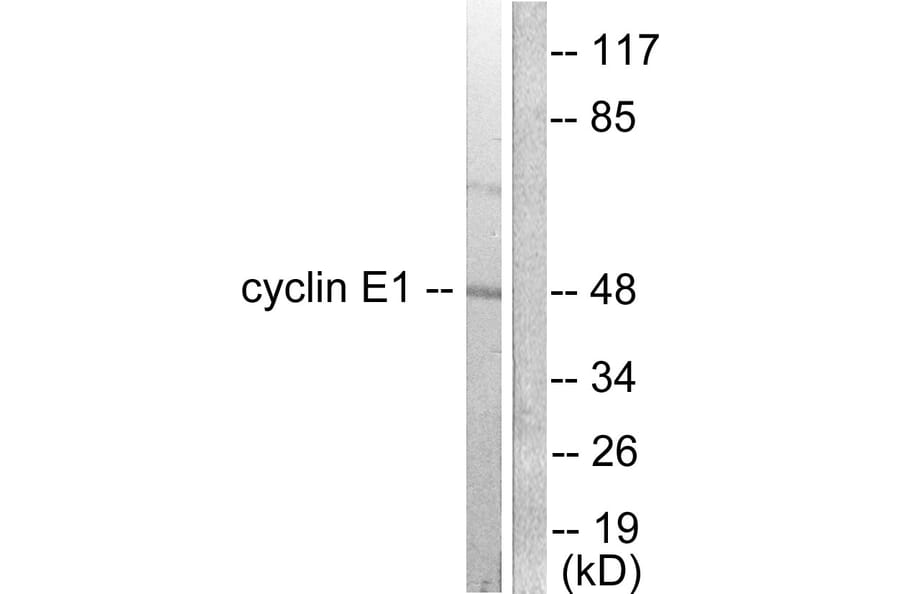
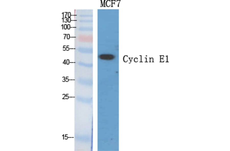
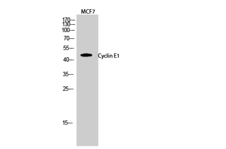
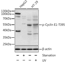
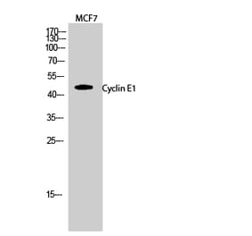
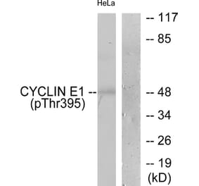
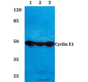
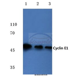
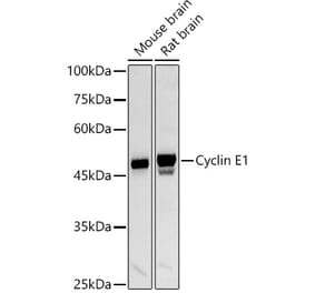
Western blot analysis of lysates from K562 cells using Anti-Cyclin E1 Antibody. The right hand lane represents a negative control, where the antibody is blocked by the immunising peptide.
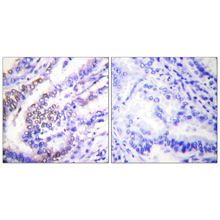
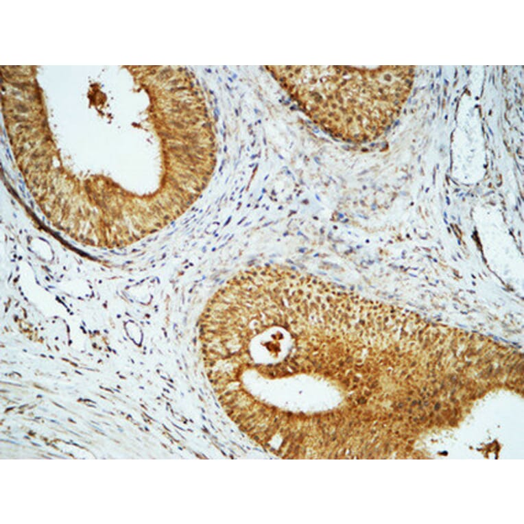
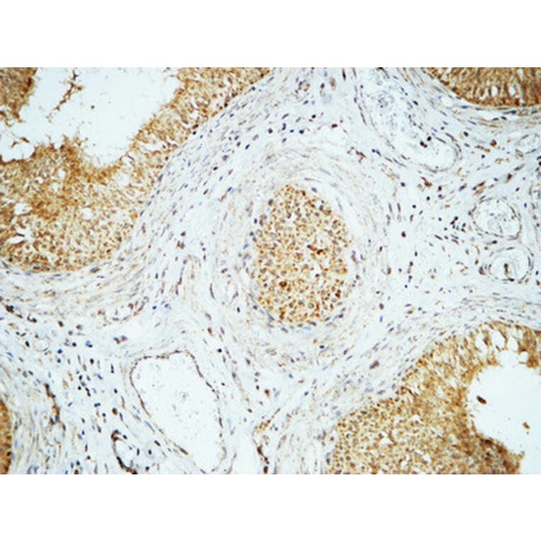
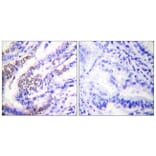
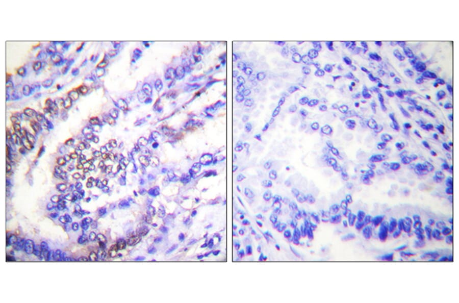
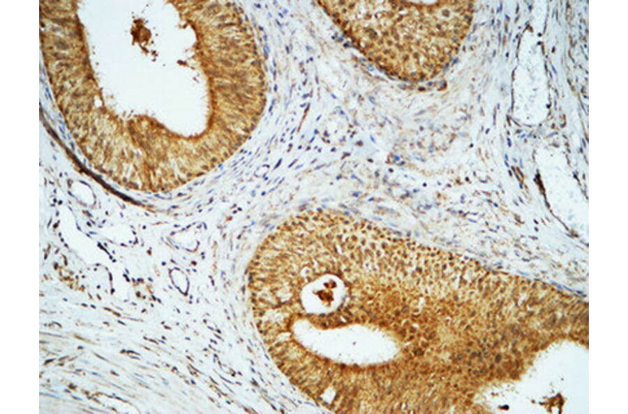
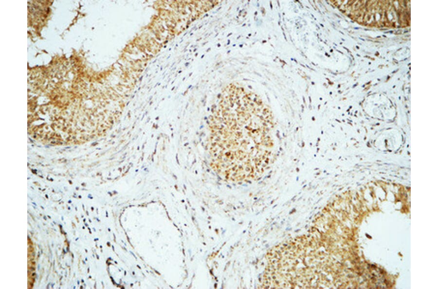
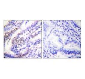
Immunohistochemical analysis of paraffin-embedded human lung carcinoma tissue using Anti-Cyclin E1 Antibody. The right hand panel represents a negative control, where the antibody was pre-incubated with the immunising peptide.
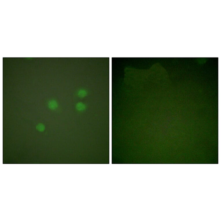
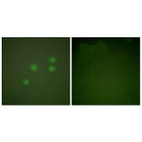
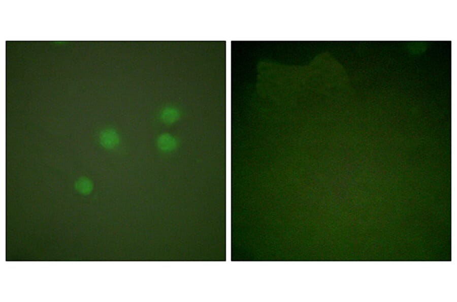
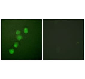
Immunofluorescence analysis of A549 cells using Anti-Cyclin E1 Antibody. The right hand panel represents a negative control, where the antibody was pre-incubated with the immunising peptide.
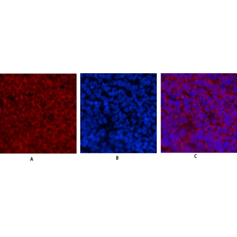
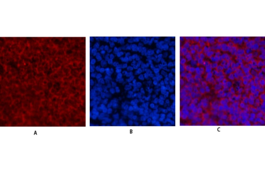
Immunofluorescence analysis of rat spleen tissue using Anti-Cyclin E1 Antibody (red) at 1:200 (4°C overnight). Cy3 labelled secondary antibody was used at 1:300 (RT 50min). Panel A: Target. Panel B: DAPI. Panel C: Merge.
Immunofluorescence analysis of rat spleen tissue using Anti-Cyclin E1 Antibody (red) at 1:200 (4°C overnight). Cy3 labelled secondary antibody was used at 1:300 (RT 50min). Panel A: Target. Panel B: DAPI. Panel C: Merge.


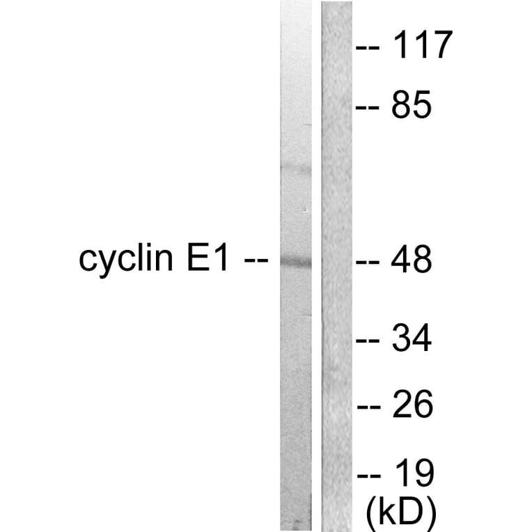
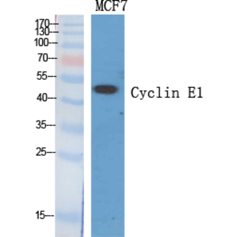
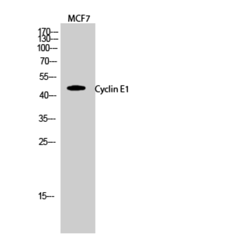

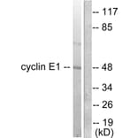
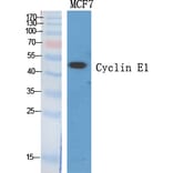


![Western Blot - Anti-Cyclin E1 Antibody [ARC51383] (A305272) - Antibodies.com](https://cdn.antibodies.com/image/catalog/305/A305272_1.jpg?profile=product_alternative)
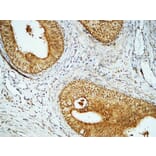
![Immunohistochemistry - Anti-Cyclin E1 Antibody [rCCNE1/4936] (A277931) - Antibodies.com](https://cdn.antibodies.com/image/catalog/277/A277931_1.jpg?profile=product_alternative)
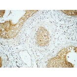
![Immunohistochemistry - Anti-Cyclin E1 Antibody [rCCNE1/4936] - BSA and Azide free (A278519) - Antibodies.com](https://cdn.antibodies.com/image/catalog/278/A278519_1.jpg?profile=product_alternative)