CD63 is expressed on activated platelets, monocytes and macrophages, and is weakly expressed on granulocytes, T cell and B cells. It is located on the basophilic granule membranes and on the plasma membranes of lymphocytes and granulocytes. CD63 is a member of the TM4 superfamily of leukocyte glycoproteins that includes CD9, CD37 and CD53, which contain four transmembrane regions. CD63 may play a role in phagocytic and intracellular lysosome-phagosome fusion events. CD63 deficiency is associated with Hermansky-Pudlak syndrome and is strongly expressed during the early stages of melanoma progression.
Applications
FUNC, IP, Flow Cytometry, IF, IHC-P
Positive Control
HL60, THP-1 or NIH/3T3 cells. Human spleen, melanoma or lymphoma.
Dilutions
Functional studies, IP: 1-2 µg per 100-500ug of total protein (1ml of cell lysate), Flow Cytometry: 1-2 µg/million cells, IF: 1-2 µg/ml, IHC: 1-2 µg/ml
Reactivity
Human
Immunogen
Recombinant fragment (around Amino Acids 100-200) of human CD63 protein. The exact sequence is proprietary.
Host
Mouse
Clonality
Monoclonal
Clone ID
LAMP3/7369
Isotype
IgG2
Light Chains
kappa
Conjugate
Unconjugated
Purification
Protein A/G chromatography.
Concentration
200 µg/ml
Molecular Weight
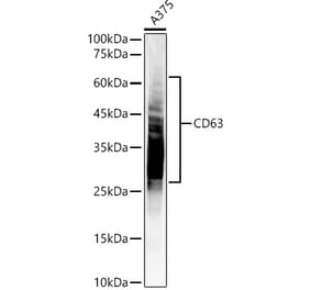
26 kDa (core protein) / 30-60 kDa (glycosylated)
Product Form
Liquid
Formulation
Supplied in 10mM Phosphate Buffered Saline with 0.05% BSA and 0.05% Sodium Azide.
Storage
Shipped at 4°C. Upon delivery aliquot and store at -20°C. Avoid freeze / thaw cycles.
Synonyms
CD63 antigen, Granulophysin, LAMP-3, Limp1, Lysosomal-associated membrane protein 3, Lysosome integral membrane protein 1, Melanoma-associated antigen ME491, MLA1, OMA81H, TSPAN30


![Flow Cytometry - Anti-CD63 Antibody [MEM-259] (A85975) - Antibodies.com](https://cdn.antibodies.com/image/catalog/85/A85976_346.jpg?profile=product_alternative)

![Western Blot - Anti-CD63 Antibody [ARC51703] (A305333) - Antibodies.com](https://cdn.antibodies.com/image/catalog/305/A305333_1.jpg?profile=product_alternative)
![Immunohistochemistry - Anti-CD63 Antibody [MX-49.129.5] (A250748) - Antibodies.com](https://cdn.antibodies.com/image/catalog/250/A250748_1.jpg?profile=product_alternative)
![Immunohistochemistry - Anti-CD63 Antibody [MX-49.129.5] - BSA and Azide free (A253928) - Antibodies.com](https://cdn.antibodies.com/image/catalog/253/A253928_1.jpg?profile=product_alternative)
![Immunohistochemistry - Anti-CD63 Antibody [NKI/C3] (A250746) - Antibodies.com](https://cdn.antibodies.com/image/catalog/250/A250746_1.jpg?profile=product_alternative)
![Immunohistochemistry - Anti-CD63 Antibody [NKI/C3] - BSA and Azide free (A253926) - Antibodies.com](https://cdn.antibodies.com/image/catalog/253/A253926_1.jpg?profile=product_alternative)
![Immunohistochemistry - Anti-CD63 Antibody [rMX-49.129.5] (A250752) - Antibodies.com](https://cdn.antibodies.com/image/catalog/250/A250752_1.jpg?profile=product_alternative)
![Immunohistochemistry - Anti-CD63 Antibody [LAMP3/2881] - BSA and Azide free (A253924) - Antibodies.com](https://cdn.antibodies.com/image/catalog/253/A253924_1.jpg?profile=product_alternative)
![Immunohistochemistry - Anti-CD63 Antibody [LAMP3/2789] (A250754) - Antibodies.com](https://cdn.antibodies.com/image/catalog/250/A250754_1.jpg?profile=product_alternative)
![Immunohistochemistry - Anti-CD63 Antibody [LAMP3/2789] - BSA and Azide free (A253934) - Antibodies.com](https://cdn.antibodies.com/image/catalog/253/A253934_1.jpg?profile=product_alternative)