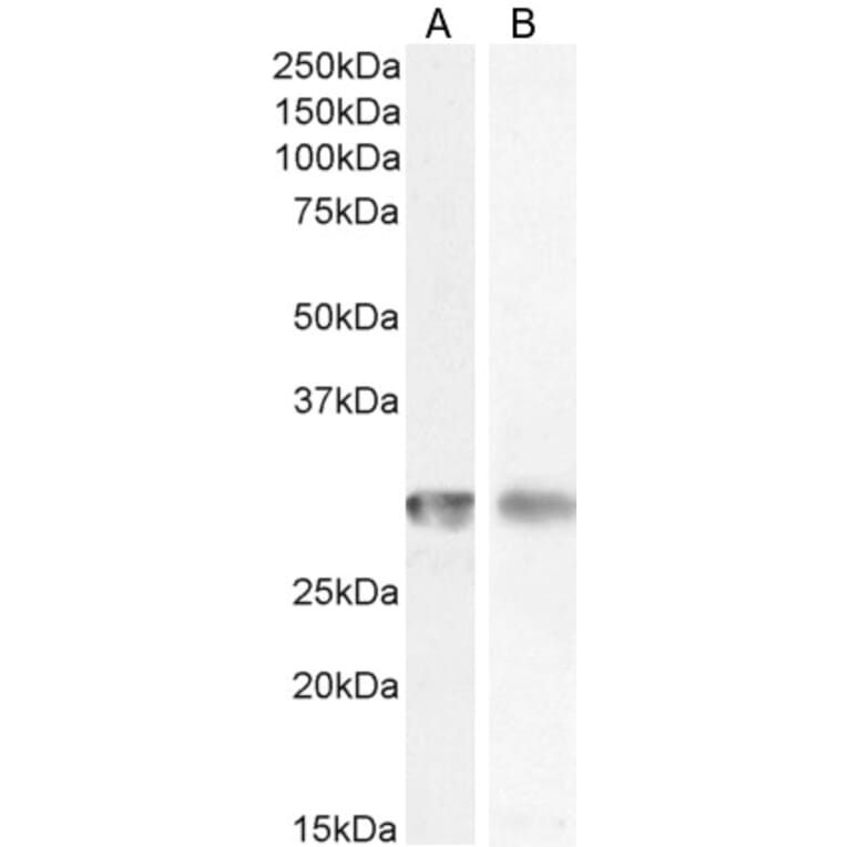
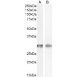
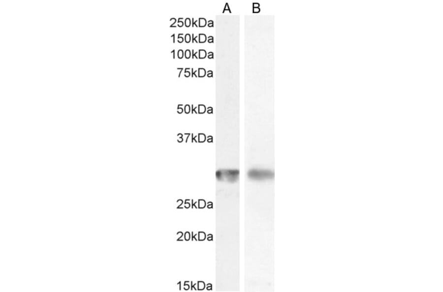
CD4 expression in Human Spleen (A) and Bone Marrow (B) lysate analyzed by western blot. Cells were lysed in RIPA buffer and 35µg protein was run per lane. Primary incubation was performed with Anti-CD4 Antibody (A82715) at 1µg/ml (A) or 0.5µg/ml (B) and detected by chemiluminescence.
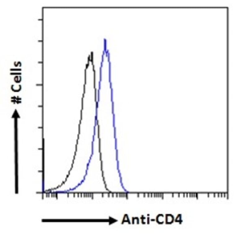
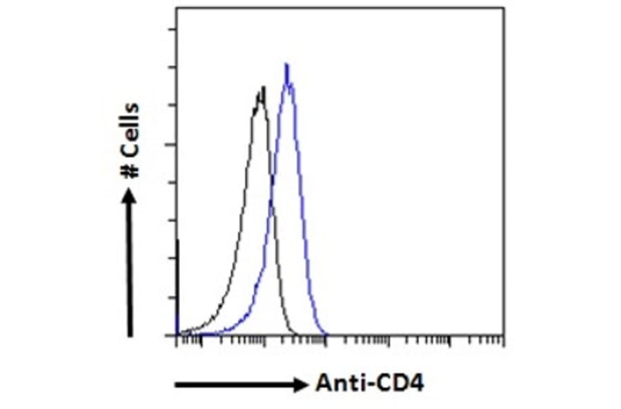
CD4 expression in Jurkat cells (blue line) analyzed by flow cytometry. Cells were fixed in PFA and permeabilized with 0.5% Triton. Staining was performed with Anti-CD4 Antibody (A82715) at 10µg/ml for 1 hour and Alexa Fluor 488 secondary antibody at 1µg/ml. Negative Control: Goat IgG Isotype Control (black line) followed by Alexa Fluor 488 secondary antibody.
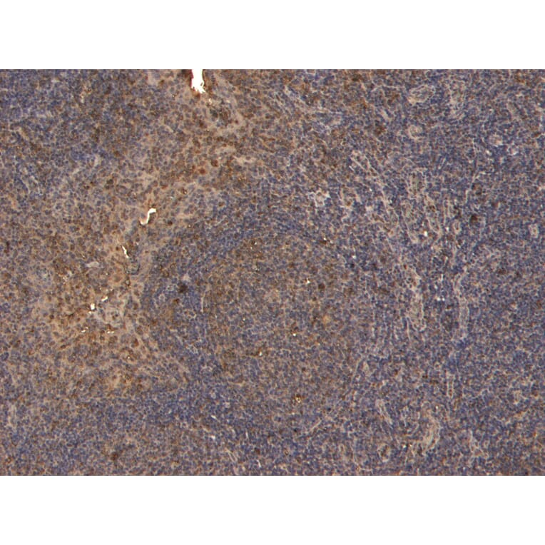
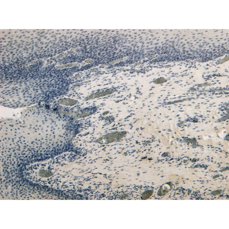
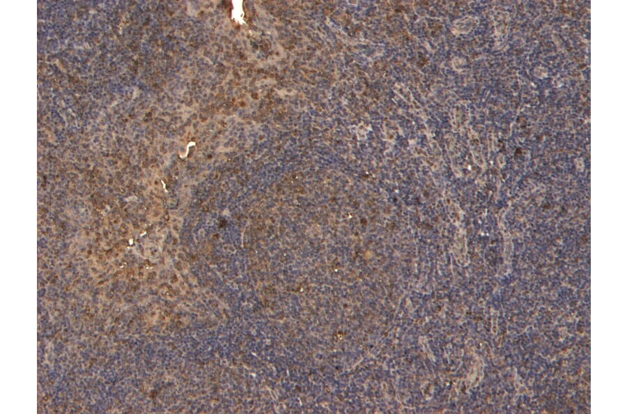
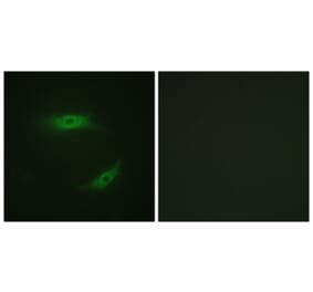
CD4 expression in Human Tonsil analyzed by immunohistochemistry. Tissue was paraffin-embedded, and antigen retrieval was achieved by heating in citrate buffer, pH 6. Staining was performed with Anti-CD4 Antibody (A82715) at 4µg/ml and revealed with horseradish peroxidase (HRP).
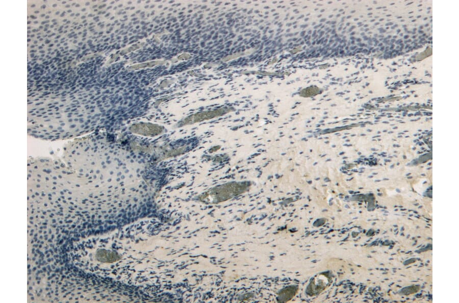
Negative control for CD4 expression in Human Tonsil analyzed by immunohistochemistry. Tissue was paraffin-embedded, and staining procedure was performed in the absence of primary antibody.
Publishing research using Anti-CD4 Antibody (A82715)? Please let us know so that we can list the citation on this page.
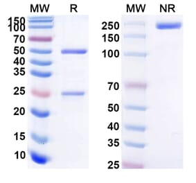
Alternative products to Anti-CD4 Antibody (A82715)


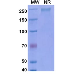
![SDS-PAGE - Anti-CD4 Antibody [M-T413] Biosimilar - BSA and Azide free (A339489) - Antibodies.com](https://cdn.antibodies.com/image/catalog/339/A339489_1.jpg?profile=product_alternative)
![SDS-PAGE - Anti-CD4 Antibody [IT128] Biosimilar - BSA and Azide free (A339490) - Antibodies.com](https://cdn.antibodies.com/image/catalog/339/A339490_1.jpg?profile=product_alternative)
![SDS-PAGE - Anti-CD4 Antibody [TRX1] Biosimilar - BSA and Azide free (A339491) - Antibodies.com](https://cdn.antibodies.com/image/catalog/339/A339491_1.jpg?profile=product_alternative)