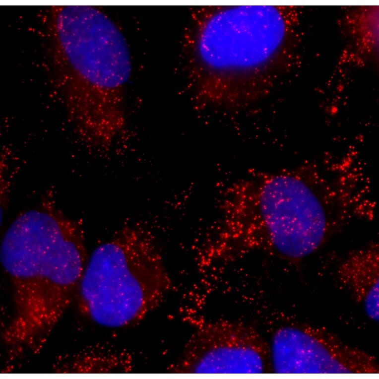
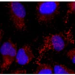
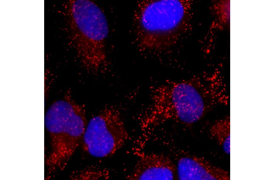
Immunofluorescent analysis of HeLa cell culture stained with Anti-Catalase Antibody [6H14] (A333282) at a dilution of 1:200 (red). Nuclei were stained with Hoechst (blue). Anti-Catalase Antibody [6H14] (A333282) antibody reveals visicular staining of catalase protein in peroxisomes in the cytoplasm.
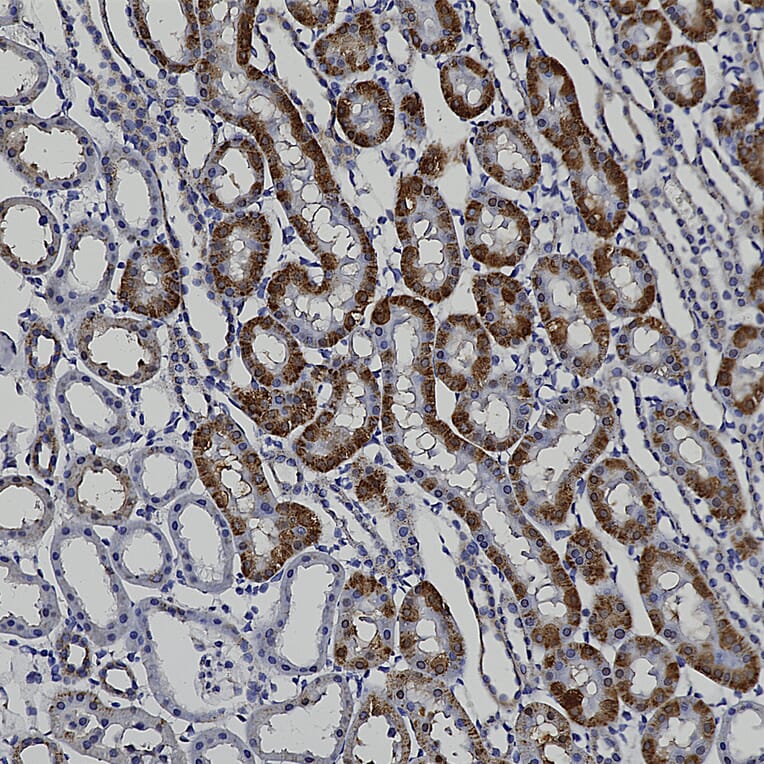
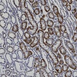
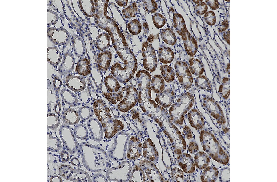
Immunohistochemistry analysis of a formalin fixed paraffin embedded rat kidney section with Anti-Catalase Antibody [6H14] (A333282) at a dilution of 1:1,000 detected with DAB (brown) using the Vector Labs ImmPRESS method and reagents with citra buffer retrieval. Counterstained with Hematoxylin (blue). The Anti-Catalase Antibody [6H14] (A333282) strongly labels the peroxisomes in the cytoplasm of kidney tubule cells. Note: this antibody performs well in testing with 4% paraformaldehyde and standard neutral buffered formalin on paraffin sections of fixed mouse, rat, and human tissue.
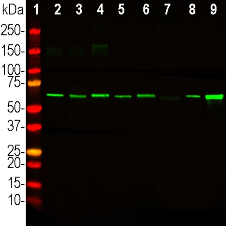
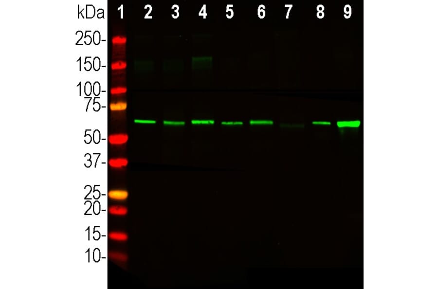
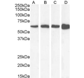
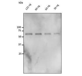
Western Blot - Anti-Catalase Antibody [6H14] (A333282)
Western blot analysis of cell lysates from various cell lines using Anti-Catalase Antibody [6H14] (A333282) at a dilution of 1:500 (green). The lanes contain: [Lane 1] protein standard (red), [Lane 2] HeLa, [Lane 3] SH-SY5Y, [Lane 4] COS-1, [Lane 5] NBL6, [Lane 6] A72, [Lane 7] NIH/3T3, [Lane 8] PC12, and [Lane 9] C6. Strong band at about 60kDa corresponds to catalase protein.
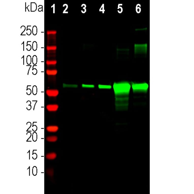
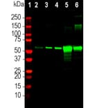
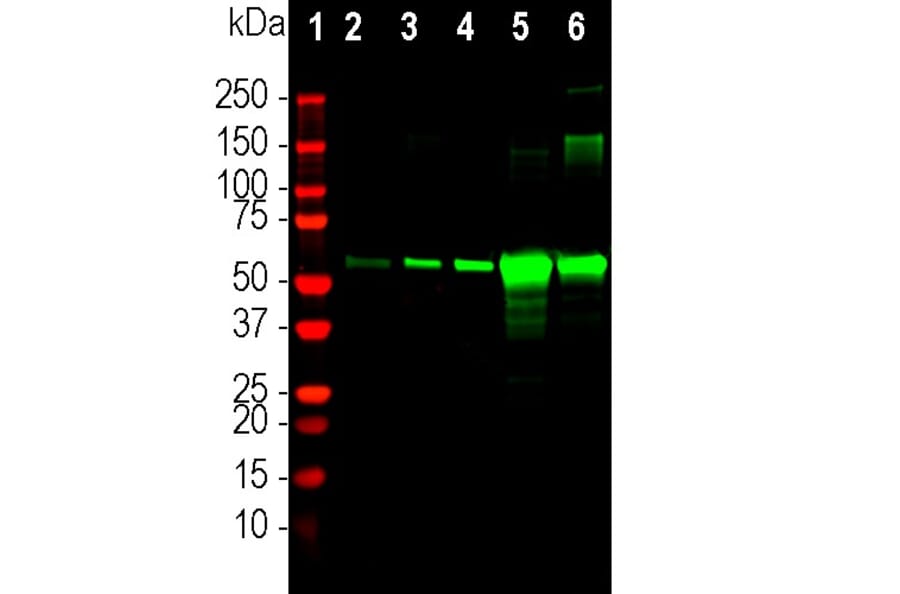
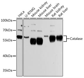
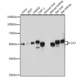
Western Blot - Anti-Catalase Antibody [6H14] (A333282)
Western blot analysis of mouse different tissue lysates using Anti-Catalase Antibody [6H14] (A333282) at a dilution of 1:500 (green). The lanes contain: [Lane 1] protein standard (red), mouse tissue:, [Lane 2] brain, [Lane 3] lungs, [Lane 4] heart, [Lane 5] liver, and [Lane 6] kidney. Strong band at about 60kDa corresponds to catalase protein. The protein is particularly abundant in kidney and especially liver.
Publishing research using Anti-Catalase Antibody [6H14] (A333282)? Please let us know so that we can list the citation on this page.
Alternative products to Anti-Catalase Antibody [6H14] (A333282)