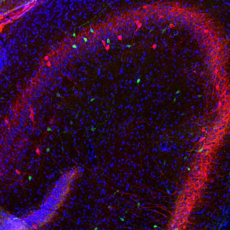
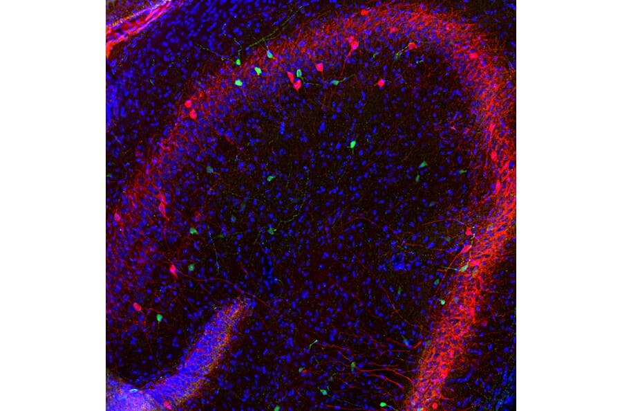
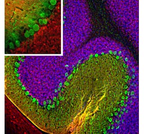
Immunofluorescent analysis of rat hippocampus section stained with Anti-Calretinin Antibody (A104312), at a dilution of 1:1,000, in green, and co-stained with Anti-Parvalbumin Antibody [3C9] (A85317), at a dilution of 1:1,000, in red. Following transcardial perfusion of rat with 4% paraformaldehyde, brain was post fixed for 24 hours, cut to 45µM, and free-floating sections were stained with the above antibodies. The Anti-Calretinin Antibody (A104312) and Anti-Parvalbumin Antibody [3C9] (A85317) label different classes of interneurons.
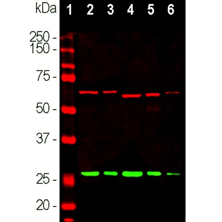
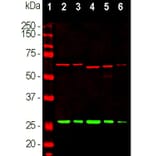
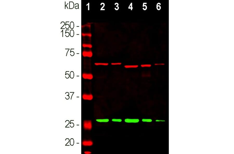
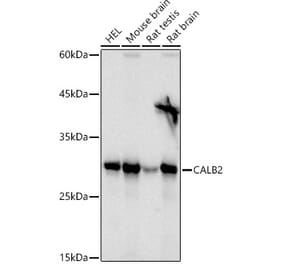
Western blot analysis of different tissue lysates using Anti-Calretinin Antibody (A104312), at a dilution of 1:1,000, in green. The lanes contain: [Lane 1] protein standard (red), [Lane 2] rat brain, [Lane 3] rat spinal cord, [Lane 4] mouse brain, [Lane 5] mouse spinal cord, and [Lane 6] cow spinal cord. A band at 29kDa corresponds to Calretinin protein. The same blot was simultaneously probed with Anti-alpha Internexin Antibody [2E3] (A85448), at a dilution of 1:10,000, in red, that reveals the alpha Internexin protein with apparent molecular weight of 66kDa.
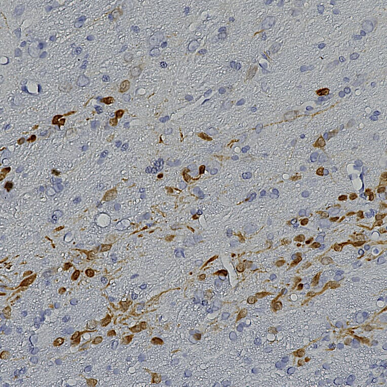
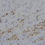
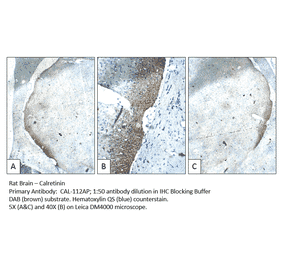
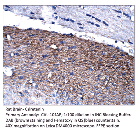
Immunohistochemistry analysis of a formalin fixed paraffin embedded rat brain section with Anti-Calretinin Antibody (A104312) at a dilution of 1:2,000 detected in DAB (brown) following the ABC method. Counterstained with Hematoxylin (blue). The subfornical organ region is imaged, showing strong labeling of dorsolateral peripheral neurons (9).
Publishing research using Anti-Calretinin Antibody (A104312)? Please let us know so that we can list the citation on this page.
Alternative products to Anti-Calretinin Antibody (A104312)


![Immunofluorescence - Anti-Calretinin Antibody [6A9] (A85366) - Antibodies.com](https://cdn.antibodies.com/image/catalog/85/A85366_1.jpg?profile=product_alternative)
![Immunofluorescence - Anti-Calretinin Antibody [3G9] (A85367) - Antibodies.com](https://cdn.antibodies.com/image/catalog/85/A85367_1.jpg?profile=product_alternative)
![Immunohistochemistry - Anti-Calretinin Antibody [CALB2/2602] (A250372) - Antibodies.com](https://cdn.antibodies.com/image/catalog/250/A250372_1.jpg?profile=product_alternative)
![Immunohistochemistry - Anti-Calretinin Antibody [CALB2/2807] - BSA and Azide free (A253556) - Antibodies.com](https://cdn.antibodies.com/image/catalog/253/A253556_1.jpg?profile=product_alternative)
![Immunohistochemistry - Anti-Calretinin Antibody [CALB2/2786] - BSA and Azide free (A253555) - Antibodies.com](https://cdn.antibodies.com/image/catalog/253/A253555_1.jpg?profile=product_alternative)
![Immunohistochemistry - Anti-Calretinin Antibody [CALB2/2807] (A250376) - Antibodies.com](https://cdn.antibodies.com/image/catalog/250/A250376_1.jpg?profile=product_alternative)
![Immunohistochemistry - Anti-Calretinin Antibody [CALB2/2786] (A250375) - Antibodies.com](https://cdn.antibodies.com/image/catalog/250/A250375_1.jpg?profile=product_alternative)
![Immunohistochemistry - Anti-Calretinin Antibody [CALB2/2602] - BSA and Azide free (A253552) - Antibodies.com](https://cdn.antibodies.com/image/catalog/253/A253552_1.jpg?profile=product_alternative)