Immunofluorescence - Anti-Calretinin Antibody [6A9] (A85366)
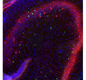
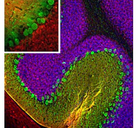
Left: Mixed neuron/glial cultures stained with Anti-Calretinin Antibody, at a dilution of 1:2,000, in red, and Anti-Vimentin Antibody (A85421 | 1:5,000, in green. Calretinin is prominently expressed in small number of interneurons, while astrocytes and fibroblasts were visualized with the Anti-Vimentin Antibody. Middle: Adult rat cortical section (45 µM; fixed by transcardial perfusion with 4% paraformaldehyde) was co-stained with Anti-Calretinin Antibody, at a dilution of 1:1,000, in red, and Anti-Calbinidin Antibody (A85365 | 1:1,000, in green. In the motor cortex, calretinin is expressed in a small population of interneurons that do not express calbindin. Because each antibody specifically labels a different population of cells exclusively, the cells are either stained with red or green. Right: Adult mouse brain hippocampal section (45 µM; fixed by transcardial perfusion with 4% paraformaldehyde) was co-stained with Anti-Calretinin Antibody, at a dilution of 1:1,000, in red, and Anti-Calbindin Antibody (A85365 | 1:1,000, in green. In the stratum radiatum of CA1 region, calretinin expresses in a small number of interneurons that do not express calbindin. As a result, our antibodies label different neurons in either red or green. Insets are high-magnification images of the boxed area in each picture. Blue is a hoechst DNA staining.


![Immunofluorescence - Anti-Calretinin Antibody [6A9] (A85366) - Antibodies.com](https://cdn.antibodies.com/image/catalog/85/A85366_1.jpg?profile=product_top)
![Immunofluorescence - Anti-Calretinin Antibody [6A9] (A85366) - Antibodies.com](https://cdn.antibodies.com/image/catalog/85/A85366_2.jpg?profile=product_top)
![Western Blot - Anti-Calretinin Antibody [6A9] (A85366) - Antibodies.com](https://cdn.antibodies.com/image/catalog/85/A85366_3.jpg?profile=product_top)
![Western Blot - Anti-Calretinin Antibody [6A9] (A85366) - Antibodies.com](https://cdn.antibodies.com/image/catalog/85/A85366_5.jpg?profile=product_top)
![Immunofluorescence - Anti-Calretinin Antibody [6A9] (A85366) - Antibodies.com](https://cdn.antibodies.com/image/catalog/85/A85366_1.jpg?profile=product_top_thumb)
![Immunofluorescence - Anti-Calretinin Antibody [6A9] (A85366) - Antibodies.com](https://cdn.antibodies.com/image/catalog/85/A85366_2.jpg?profile=product_top_thumb)
![Western Blot - Anti-Calretinin Antibody [6A9] (A85366) - Antibodies.com](https://cdn.antibodies.com/image/catalog/85/A85366_3.jpg?profile=product_top_thumb)
![Western Blot - Anti-Calretinin Antibody [6A9] (A85366) - Antibodies.com](https://cdn.antibodies.com/image/catalog/85/A85366_5.jpg?profile=product_top_thumb)
![Immunofluorescence - Anti-Calretinin Antibody [6A9] (A85366) - Antibodies.com](https://cdn.antibodies.com/image/catalog/85/A85366_1.jpg?profile=product_image)
![Immunofluorescence - Anti-Calretinin Antibody [6A9] (A85366) - Antibodies.com](https://cdn.antibodies.com/image/catalog/85/A85366_2.jpg?profile=product_image)
![Western Blot - Anti-Calretinin Antibody [6A9] (A85366) - Antibodies.com](https://cdn.antibodies.com/image/catalog/85/A85366_3.jpg?profile=product_image)
![Western Blot - Anti-Calretinin Antibody [6A9] (A85366) - Antibodies.com](https://cdn.antibodies.com/image/catalog/85/A85366_5.jpg?profile=product_image)
![Immunohistochemistry - Anti-Calretinin Antibody [CALB2/2786] (A250375) - Antibodies.com](https://cdn.antibodies.com/image/catalog/250/A250375_1.jpg?profile=product_alternative)
![Immunohistochemistry - Anti-Calretinin Antibody [CALB2/2602] - BSA and Azide free (A253552) - Antibodies.com](https://cdn.antibodies.com/image/catalog/253/A253552_1.jpg?profile=product_alternative)
![Immunohistochemistry - Anti-Calretinin Antibody [CALB2/2685] (A250374) - Antibodies.com](https://cdn.antibodies.com/image/catalog/250/A250374_1.jpg?profile=product_alternative)
![Immunohistochemistry - Anti-Calretinin Antibody [CALB2/2685] - BSA and Azide free (A253554) - Antibodies.com](https://cdn.antibodies.com/image/catalog/253/A253554_1.jpg?profile=product_alternative)
![Immunofluorescence - Anti-Calretinin Antibody [3G9] (A85367) - Antibodies.com](https://cdn.antibodies.com/image/catalog/85/A85367_1.jpg?profile=product_alternative)
![Immunohistochemistry - Anti-Calretinin Antibody [CALB2/2602] (A250372) - Antibodies.com](https://cdn.antibodies.com/image/catalog/250/A250372_1.jpg?profile=product_alternative)
![Immunohistochemistry - Anti-Calretinin Antibody [CALB2/2807] - BSA and Azide free (A253556) - Antibodies.com](https://cdn.antibodies.com/image/catalog/253/A253556_1.jpg?profile=product_alternative)
![Immunohistochemistry - Anti-Calretinin Antibody [CALB2/2786] - BSA and Azide free (A253555) - Antibodies.com](https://cdn.antibodies.com/image/catalog/253/A253555_1.jpg?profile=product_alternative)
![Immunohistochemistry - Anti-Calretinin Antibody [CALB2/2807] (A250376) - Antibodies.com](https://cdn.antibodies.com/image/catalog/250/A250376_1.jpg?profile=product_alternative)