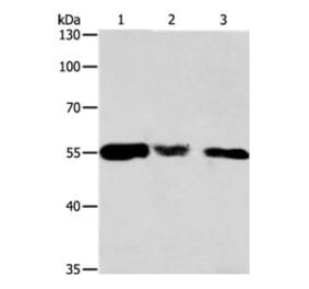
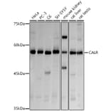
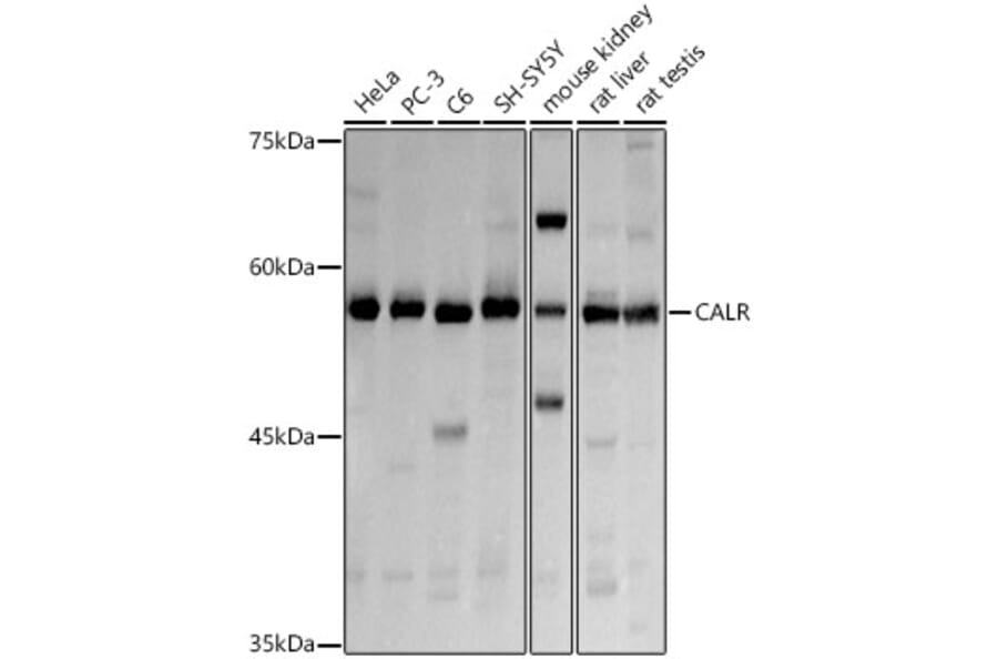
Western Blot - Anti-Calreticulin Antibody (A13022)
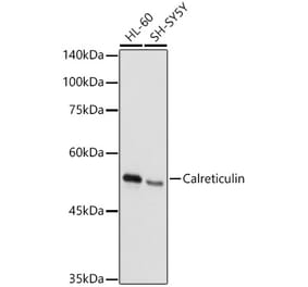
Western blot analysis of extracts of various cell lines, using Anti-Calreticulin Antibody (A13022) at 1:3,000 dilution. The secondary antibody was Goat Anti-Rabbit IgG H&L Antibody (HRP) at 1:10,000 dilution. Lysates/proteins were present at 25µg per lane. The blocking buffer used was 3% non-fat dry milk in TBST. Detection was with a ECL Basic Kit. Exposure time: 1s.
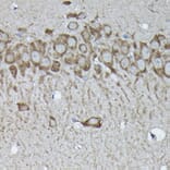
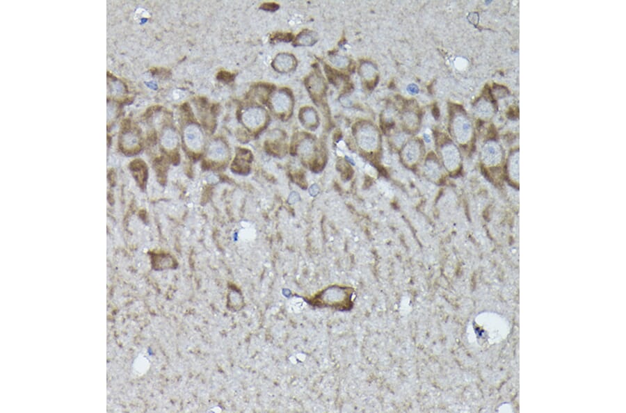
Immunohistochemistry analysis of paraffin-embedded rat brain using Anti-Calreticulin Antibody (A13022) at a dilution of 1:100 (40x lens). Perform high pressure antigen retrieval with 10 mM citrate buffer pH 6.0 before commencing with IHC staining protocol.
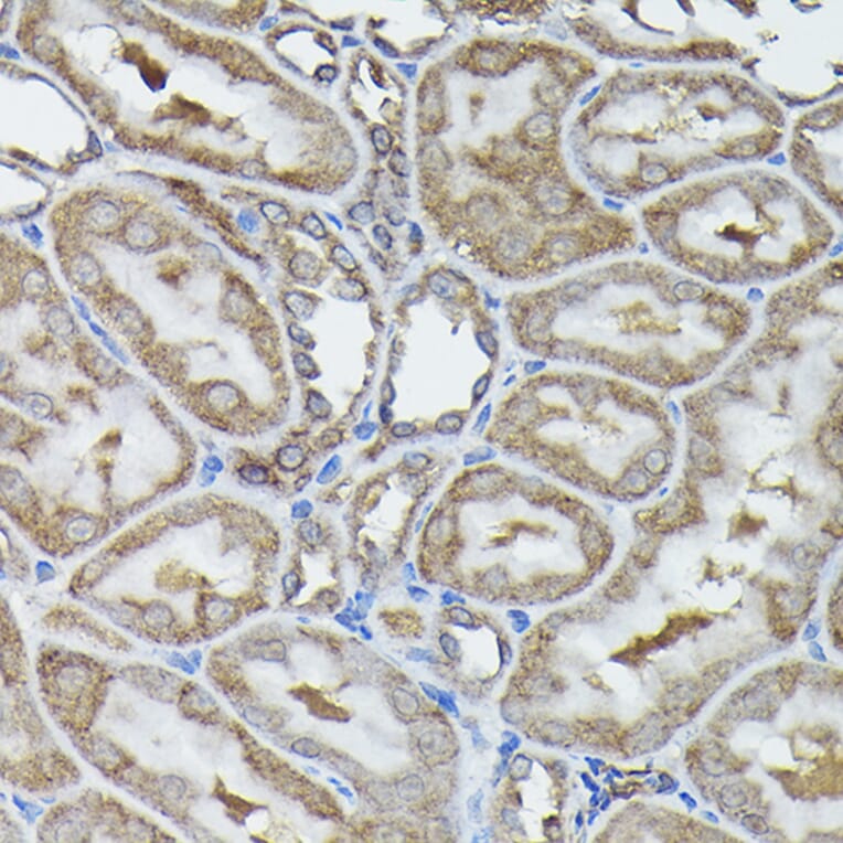
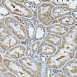
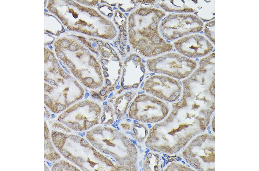
Immunohistochemistry analysis of paraffin-embedded rat kidney using Anti-Calreticulin Antibody (A13022) at a dilution of 1:100 (40x lens). Perform high pressure antigen retrieval with 10 mM citrate buffer pH 6.0 before commencing with IHC staining protocol.
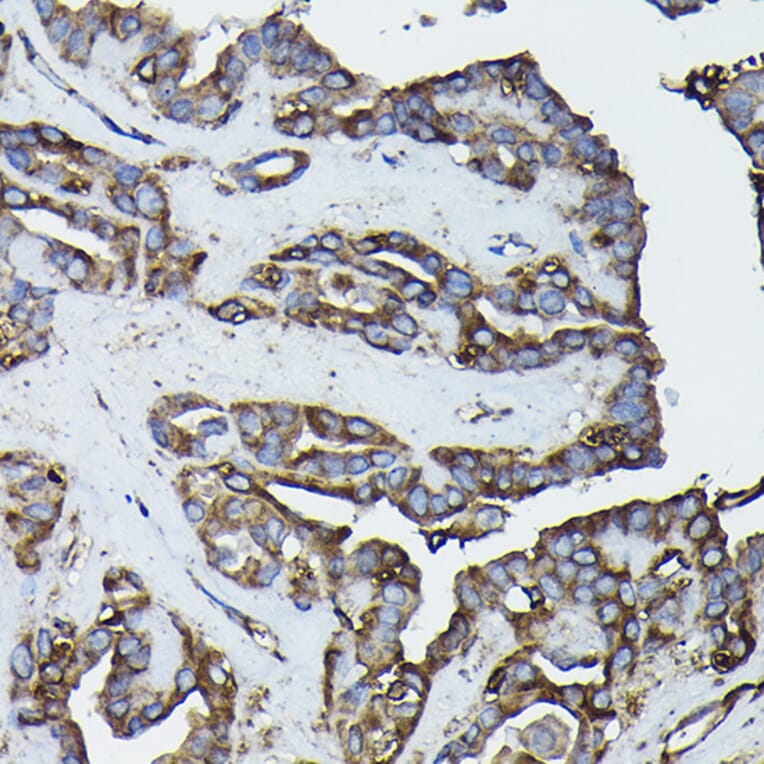
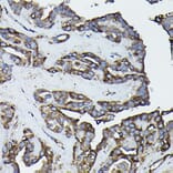
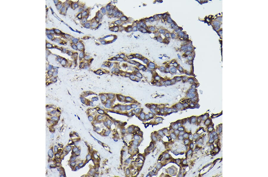
Immunohistochemistry analysis of paraffin-embedded human thyroid cancer using Anti-Calreticulin Antibody (A13022) at a dilution of 1:100 (40x lens). Perform high pressure antigen retrieval with 10 mM citrate buffer pH 6.0 before commencing with IHC staining protocol.
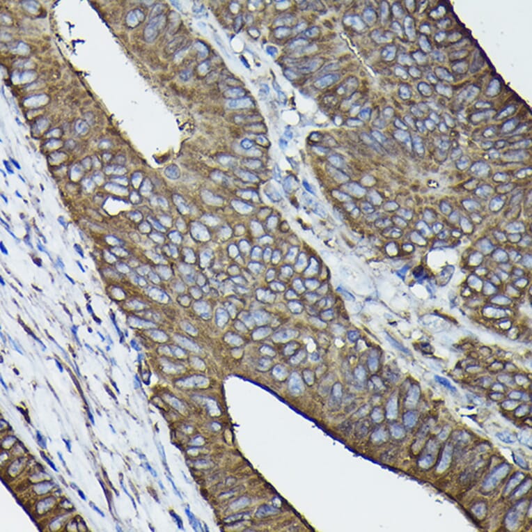
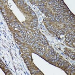
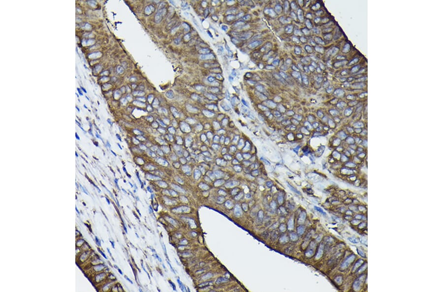
Immunohistochemistry analysis of paraffin-embedded human colon carcinoma tissue using Anti-Calreticulin Antibody (A13022) at a dilution of 1:100 (40x lens). Perform high pressure antigen retrieval with 10 mM citrate buffer pH 6.0 before commencing with IHC staining protocol.
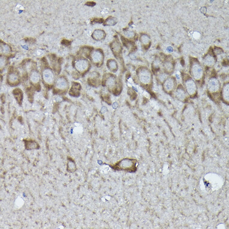
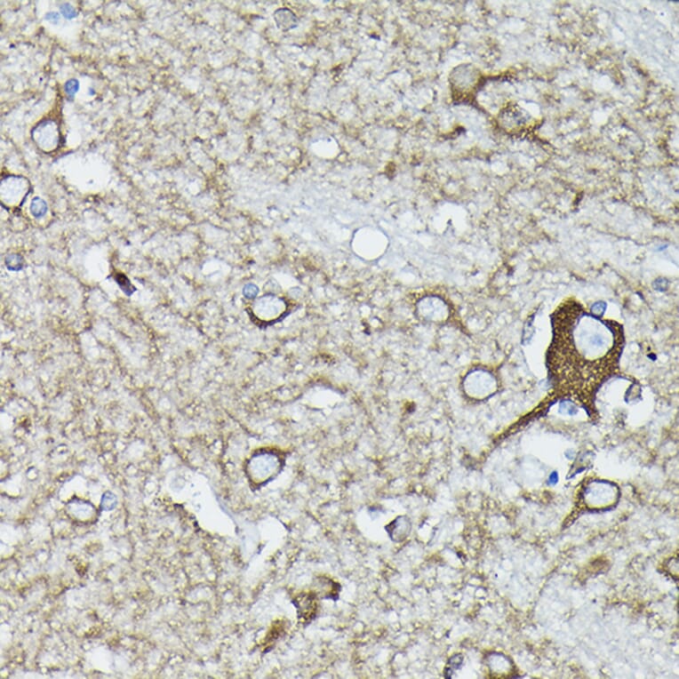
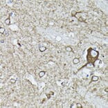
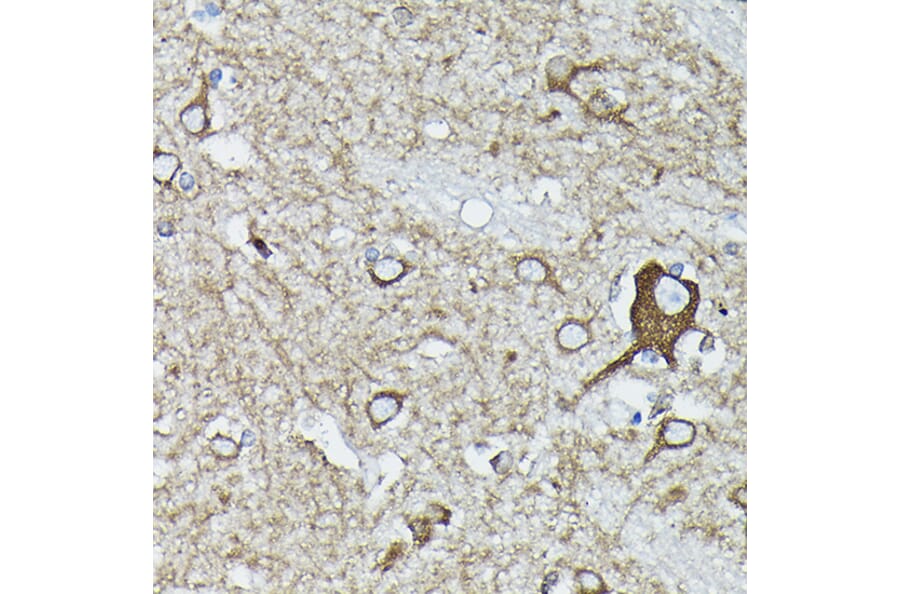
Immunohistochemistry analysis of paraffin-embedded human brain tissue using Anti-Calreticulin Antibody (A13022) at a dilution of 1:100 (40x lens). Perform high pressure antigen retrieval with 10 mM citrate buffer pH 6.0 before commencing with IHC staining protocol.
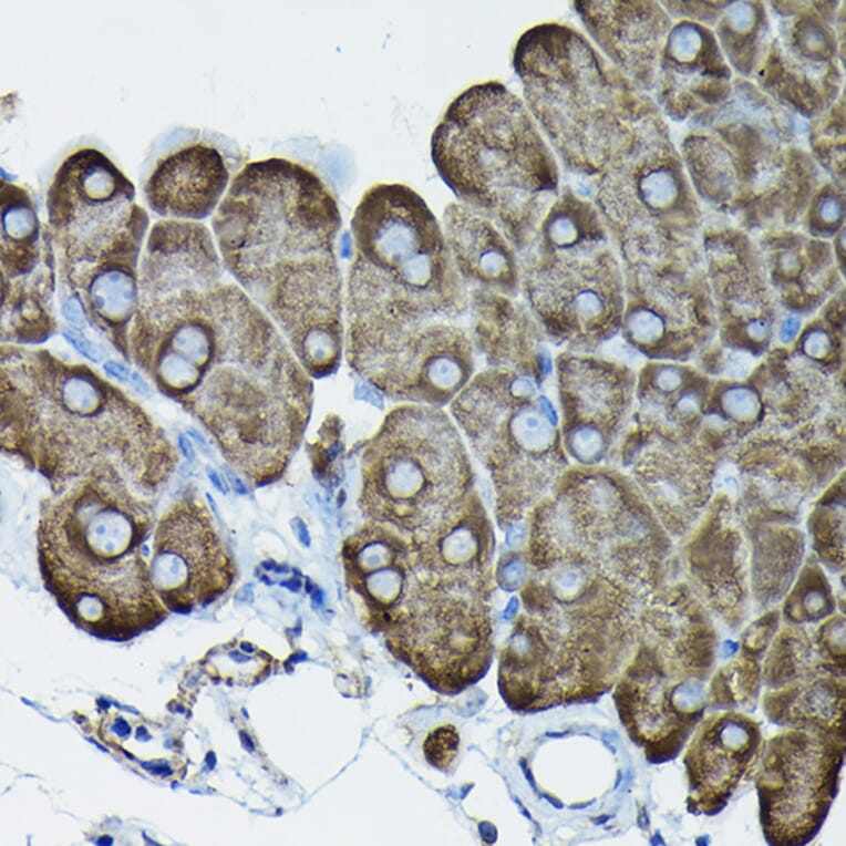
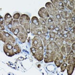
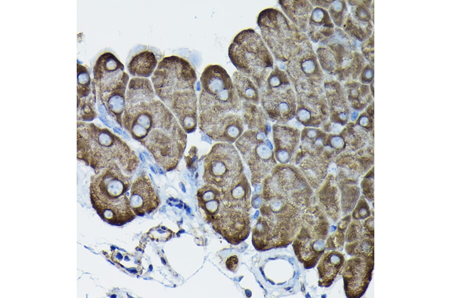
Immunohistochemistry analysis of paraffin-embedded mouse pancreas using Anti-Calreticulin Antibody (A13022) at a dilution of 1:100 (40x lens). Perform high pressure antigen retrieval with 10 mM citrate buffer pH 6.0 before commencing with IHC staining protocol.
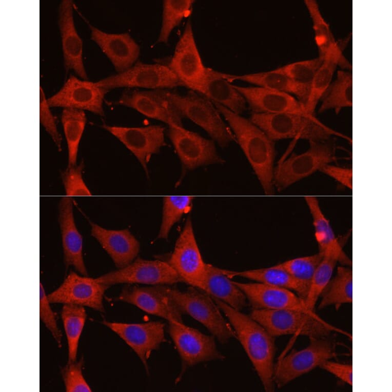
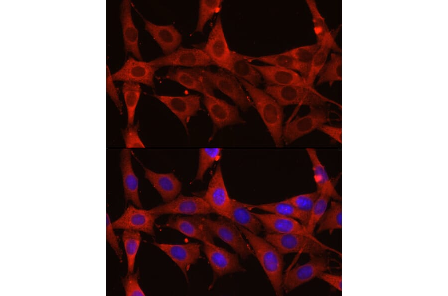
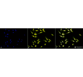
Immunofluorescence analysis of NIH/3T3 cells using Anti-Calreticulin Antibody (A13022) at a dilution of 1:200 (40x lens). DAPI was used to stain the cell nuclei (blue).
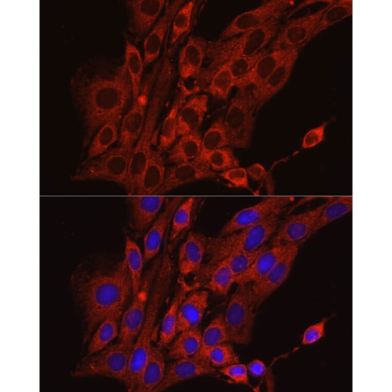
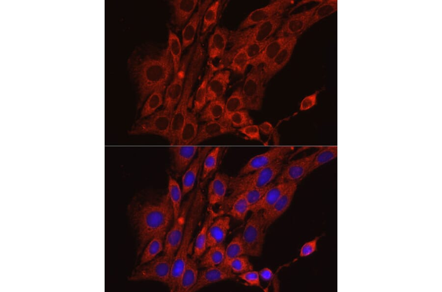
Immunofluorescence analysis of PC-12 cells using Anti-Calreticulin Antibody (A13022) at a dilution of 1:200 (40x lens). DAPI was used to stain the cell nuclei (blue).
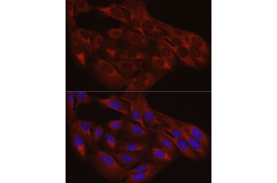
Immunofluorescence analysis of U2OS cells using Anti-Calreticulin Antibody (A13022) at a dilution of 1:200 (40x lens). DAPI was used to stain the cell nuclei (blue).


![Immunofluorescence - Anti-Calreticulin Antibody [6C6] (A85411) - Antibodies.com](https://cdn.antibodies.com/image/catalog/85/A85411_1.jpg?profile=product_alternative)