This antibody binds Calbindin cleanly but does not cross-react with the related Calretinin and Parvalbumin proteins. It is therefore ideally suited for identifying and subclassifying cortical GABAergic neurons.
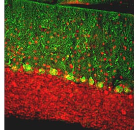
Immunofluorescent analysis of rat brain cerebellum section stained with Anti-Calbindin Antibody, at a dilution of 1:1,000, in green, and Anti-Calretinin (A85364 | 1:5,000, in red. The blue is DAPI staining of nuclear DNA. Following transcardial perfusion with 4% paraformaldehyde, brain was post fixed for 24 hours, cut to 45µM, and free-floating sections were stained with the above antibodies. Anti-Calbindin Antibody prominently labels the dendrites and perikarya of Purkinje cells in the molecular layer of cerebellum. In contrast the Anti-Calretinin Antibody stains granule cells, in the granualar layer, and their processes in the molecular layer.
Rat brain cerebellum (Middle) and cortex (Right) sections (45 µM; fixed by transcardial perfusion with 4% paraformaldehyde) were co-stained with Anti-Calbindin Antibody (red) and Anti-MeCP2 Antibody (A85427 | green). Calbindin is predominantly expressed in the dendrites and perikarya of Purkinje cells in the molecular layer of cerebellum, and selectively expressed in certain type of interneurons (calbindin-positive interneuron) in the cortex. MeCP2 is universally expressed in the nuclei of almost all neurons. As a result, calbindin-expressing cell is strongly labeled with red in soma, but the nucleus appears to be yellow. Blue is DAPI nucleus staining. Insets are high magnification images of the boxed areas.
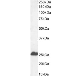
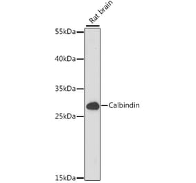
Western Blot - Anti-Calbindin Antibody [4H7] (A85360)
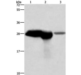
Western blot analysis of different neuronal tissue lysates using Anti-Calbindin Antibody, at a dilution of 1:2,000): [Lane 1] protein standard, [Lane 2] rat cerebellum, [Lane 3] pig hippocampus, and [Lane 4] cow cerebellum. Bands at ~25 kDa corresponds to the calbindin protein, heavily expressed in the cerebellum but a very minor component of hippocampus.
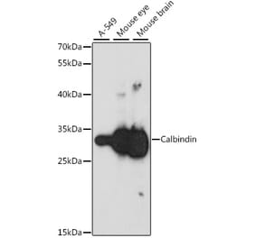
Western Blot - Anti-Calbindin Antibody [4H7] (A85360)
Western blot analysis of Anti-Calbindin Antibody. Blots of cow cerebellum lysate were probed with Anti-Calbindin Antibody. The antibody binds strongly and specifically to the calbindin band at 28 kDa.
Immunofluorescent analysis of rat cerebellum section stained with Anti-Calretinin Antibody (A104325), at a dilution of 1:5,000, in red, and co-stained with Anti-Calbindin Antibody [4H7] (A85360), at a dilution of 1:1,000, in green. Following transcardial perfusion of rat with 4% paraformaldehyde, brain was post fixed for 24 hours, cut to 45µM, and free-floating sections were stained with the above antibodies. Anti-Calretinin Antibody (A104325) stains interneurons predominantly in the molecular layer, while the Anti-Calbindin Antibody [4H7] (A85360) strongly labels the dendrites and perikarya of Purkinje cells in the molecular layer of the cerebellum.
Immunofluorescent analysis of rat brain cerebellum section stained with Anti-Myelin Basic Protein Antibody (A85321) at a dilution of 1:5,000 (red) and costained with Anti-Calbindin Antibody [4H7] (A85360) at a dilution of 1:5,000 (green). Nuclei were stained with Hoechst (blue). This antibody stains myelin sheath around axons, while the calbindin antibody prominently labels dendrites and perikarya of Purkinje cells in the molecular layer of the cerebellum.
Immunofluorescent analysis of rat brain cerebellum section stained with Anti-Myelin Basic Protein Antibody (A85321) at a dilution of 1:5,000 (red) and costained with Anti-Calbindin Antibody [4H7] (A85360) at a dilution of 1:5,000 (green). This is a higher magnification view of the specimen shown on the product home page. Nuclei were stained with Hoechst (blue). This antibody stains myelin sheath around axons, while the calbindin antibody prominently labels dendrites and perikarya of Purkinje cells in the molecular layer of the cerebellum.
Immunohistochemistry analysis of a formalin fixed paraffin embedded human cerebellum section with Anti-Calbindin Antibody [4H7] (A85360) at a dilution of 1:2,000 detected with DAB (brown) using the Vector Elite ABC-HRP detection and reagents with citra buffer retrieval. Counterstained with Hematoxylin (blue). The Anti-Calbindin Antibody [4H7] (A85360) strongly labels the cytoplasm of Purkinje cells and their dendrites. Note: this antibody performs well in testing with both 4% PFA and standard NBF fixed rat and human tissues.


![Immunofluorescence - Anti-Calbindin Antibody [4H7] (A85360) - Antibodies.com](https://cdn.antibodies.com/image/catalog/85/A85360_1.jpg?profile=product_top)
![Immunofluorescence - Anti-Calbindin Antibody [4H7] (A85360) - Antibodies.com](https://cdn.antibodies.com/image/catalog/85/A85360_2.jpg?profile=product_top)
![Western Blot - Anti-Calbindin Antibody [4H7] (A85360) - Antibodies.com](https://cdn.antibodies.com/image/catalog/85/A85360_3.jpg?profile=product_top)
![Western Blot - Anti-Calbindin Antibody [4H7] (A85360) - Antibodies.com](https://cdn.antibodies.com/image/catalog/85/A85360_5.jpg?profile=product_top)
![Immunofluorescence - Anti-Calbindin Antibody [4H7] (A85360) - Antibodies.com](https://cdn.antibodies.com/image/catalog/85/A85360_6.jpg?profile=product_top)
![Immunofluorescence - Anti-Calbindin Antibody [4H7] (A85360) - Antibodies.com](https://cdn.antibodies.com/image/catalog/85/A85360_7.jpg?profile=product_top)
![Immunofluorescence - Anti-Calbindin Antibody [4H7] (A85360) - Antibodies.com](https://cdn.antibodies.com/image/catalog/85/A85360_8.jpg?profile=product_top)
![Immunohistochemistry - Anti-Calbindin Antibody [4H7] (A85360) - Antibodies.com](https://cdn.antibodies.com/image/catalog/85/A85360_9.jpg?profile=product_top)
![Immunofluorescence - Anti-Calbindin Antibody [4H7] (A85360) - Antibodies.com](https://cdn.antibodies.com/image/catalog/85/A85360_1.jpg?profile=product_top_thumb)
![Immunofluorescence - Anti-Calbindin Antibody [4H7] (A85360) - Antibodies.com](https://cdn.antibodies.com/image/catalog/85/A85360_2.jpg?profile=product_top_thumb)
![Western Blot - Anti-Calbindin Antibody [4H7] (A85360) - Antibodies.com](https://cdn.antibodies.com/image/catalog/85/A85360_3.jpg?profile=product_top_thumb)
![Western Blot - Anti-Calbindin Antibody [4H7] (A85360) - Antibodies.com](https://cdn.antibodies.com/image/catalog/85/A85360_5.jpg?profile=product_top_thumb)
![Immunofluorescence - Anti-Calbindin Antibody [4H7] (A85360) - Antibodies.com](https://cdn.antibodies.com/image/catalog/85/A85360_6.jpg?profile=product_top_thumb)
![Immunofluorescence - Anti-Calbindin Antibody [4H7] (A85360) - Antibodies.com](https://cdn.antibodies.com/image/catalog/85/A85360_7.jpg?profile=product_top_thumb)
![Immunofluorescence - Anti-Calbindin Antibody [4H7] (A85360) - Antibodies.com](https://cdn.antibodies.com/image/catalog/85/A85360_8.jpg?profile=product_top_thumb)
![Immunofluorescence - Anti-Calbindin Antibody [4H7] (A85360) - Antibodies.com](https://cdn.antibodies.com/image/catalog/85/A85360_1.jpg?profile=product_image)
![Immunofluorescence - Anti-Calbindin Antibody [4H7] (A85360) - Antibodies.com](https://cdn.antibodies.com/image/catalog/85/A85360_2.jpg?profile=product_image)
![Western Blot - Anti-Calbindin Antibody [4H7] (A85360) - Antibodies.com](https://cdn.antibodies.com/image/catalog/85/A85360_3.jpg?profile=product_image)
![Western Blot - Anti-Calbindin Antibody [4H7] (A85360) - Antibodies.com](https://cdn.antibodies.com/image/catalog/85/A85360_5.jpg?profile=product_image)
![Immunofluorescence - Anti-Calbindin Antibody [4H7] (A85360) - Antibodies.com](https://cdn.antibodies.com/image/catalog/85/A85360_6.jpg?profile=product_image)
![Immunofluorescence - Anti-Calbindin Antibody [4H7] (A85360) - Antibodies.com](https://cdn.antibodies.com/image/catalog/85/A85360_7.jpg?profile=product_image)
![Immunofluorescence - Anti-Calbindin Antibody [4H7] (A85360) - Antibodies.com](https://cdn.antibodies.com/image/catalog/85/A85360_8.jpg?profile=product_image)
![Immunohistochemistry - Anti-Calbindin Antibody [4H7] (A85360) - Antibodies.com](https://cdn.antibodies.com/image/catalog/85/A85360_9.jpg?profile=product_image)
![Immunofluorescence - Anti-Calbindin Antibody [5A9] (A85362) - Antibodies.com](https://cdn.antibodies.com/image/catalog/85/A85362_1.jpg?profile=product_alternative)
![Immunohistochemistry - Anti-Calbindin Antibody [CALB1/3333] (A250371) - Antibodies.com](https://cdn.antibodies.com/image/catalog/250/A250371_1.jpg?profile=product_alternative)
![Immunohistochemistry - Anti-Calbindin Antibody [CALB1/3333] - BSA and Azide free (A253551) - Antibodies.com](https://cdn.antibodies.com/image/catalog/253/A253551_1.jpg?profile=product_alternative)
![Immunohistochemistry - Anti-Calbindin Antibody [CALB1/2782] - BSA and Azide free (A253550) - Antibodies.com](https://cdn.antibodies.com/image/catalog/253/A253550_1.jpg?profile=product_alternative)
![Immunohistochemistry - Anti-Calbindin Antibody [CALB1/2782] (A250370) - Antibodies.com](https://cdn.antibodies.com/image/catalog/250/A250370_1.jpg?profile=product_alternative)
![SDS-PAGE - Anti-Calbindin Antibody [CALB1/2364] - BSA and Azide free (A253549) - Antibodies.com](https://cdn.antibodies.com/image/catalog/253/A253549_1.jpg?profile=product_alternative)
![SDS-PAGE - Anti-Calbindin Antibody [CALB1/2364] (A250369) - Antibodies.com](https://cdn.antibodies.com/image/catalog/250/A250369_1.jpg?profile=product_alternative)
