HeLa cells stained with Anti-Beta Tubulin Antibody (green), Anti-Lamin A/C Antibody (A85443 | red) and DNA in blue. Anti-Beta Tubulin Antibody reveals strong microtubular staining in the cytoplasm of HeLa cells, while the Anti-Lamin A/C Antibody localizes in the nuclear membrane.
Immunofluorescent analysis of HeLa cells stained with Anti-Beta Tubulin Antibody, at a dilution of 1:5,000, in green, and Anti-FOX2 Antibody, at a dilution of 1:1,000, in red. Blue is DAPI staining of nuclear DNA. Anti-Beta Tubulin Antibody produces strong staining of microtubules in the cytoplasm, while the Anti-FOX2 Antibody labels the nuclei of HeLa cells.
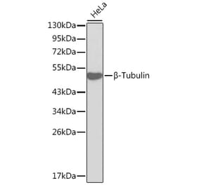
Western Blot - Anti-beta Tubulin Antibody [1B12] (A85428)
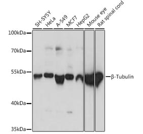
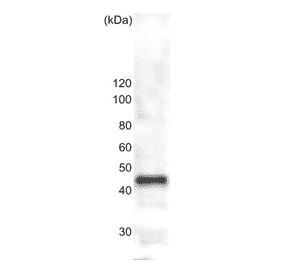
Western blot analysis of whole tissue or cell lysates using Anti-Beta Tubulin Antibody, at a dilution of 1:10,000, in green,: [Lane 1] protein standard (red), [Lane 2] adult rat brain, [Lane 3] adult mouse brain, [Lane 4] NIH-3T3 cells, [Lane 5] HEK293 cells, [Lane 6] HeLa cells, [Lane 7] SH-SY5Y cells. The strong clean band with apparent SDS-PAGE molecular weight of ~50 kDa corresponds to ß-tubulin proteins.
Immunofluorescent analysis of HeLa cells stained with Anti-MARCKS Antibody (A104335), at a dilution of 1:5,000, in red, and co-stained with Anti-beta Tubulin Antibody [1B12] (A85428), at a dilution of 1:10,000, in green. The blue is Hoechst staining of nuclear DNA. Anti-MARCKS Antibody (A104335) binds to MARCKS protein expressed in the plasma membrane and cytoplasm, while Anti-beta Tubulin Antibody [1B12] (A85428) stains cytoplasmic microtubules.
Western Blot - Anti-beta Tubulin Antibody [1B12] (A85428)
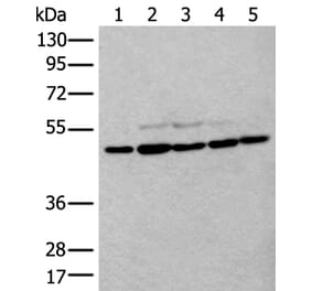
Western blot analysis of different tissue and cell lysates using Anti-DJ1 Antibody (A104338), at a dilution of 1:2,000, in green. The lanes contain: [Lane 1] protein standard, [Lane 2] rat brain, [Lane 3] mouse brain, [Lane 4] rat embryonic neuron-glial cells, [Lane 5] NIH-3T3, [Lane 6] HEK293, and [Lane 7] HeLa cells. Anti-DJ1 Antibody (A104338) detects protein with apparent molecular weight of 21kDa in all preparations with different levels of expression. The blot was simultaneously probed with Anti-beta Tubulin Antibody [1B12] (A85428), at a dilution of 1;10,000, in red, revealing a single band at ~50kDa corresponding to the beta Tubulin protein.
Western Blot - Anti-beta Tubulin Antibody [1B12] (A85428)
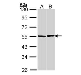
Western blot analysis of whole brain and neuron-glial cell culture lysates using Anti-FABP7 Antibody (A104339), at a dilution of 1:2,000, in green, and Anti-beta Tubulin Antibody [1B12] (A85428), at a dilution of 1:10,000, in red. The lanes contain: [Lane 1] protein standard, [Lane 2] adult rat brain, [Lane 3] E18 embryonic rat brain, [Lane 4] rat neuron-glial cell culture, and [Lane 5] adult mouse brain lysates. The band at ~14kDa corresponds to FABP7 detected only in developing tissue and cells, while the 50kDa band represents the beta Tubulin protein which is present in all preparations.
Immunofluorescent analysis of HeLa cells stained with Anti-Ki67 Antibody (A104340), at a dilution of 1:2,000, in red, and Anti-beta Tubulin Antibody [1B12] (A85428), at a dilution of 1:5,000, in green. The blue is DAPI staining of nuclear DNA. Anti-Ki67 Antibody (A104340) detects Ki67 protein predominantly expressed in nucleoli of cells in interphase and in mitotic cells Ki67 forms a coat around condensed chromosomes. Ki67 is not detected in cells in the quiescent G0 state. Anti-beta Tubulin Antibody [1B12] (A85428) produces strong staining of cytoplasmic microtubules


![Immunofluorescence - Anti-beta Tubulin Antibody [1B12] (A85428) - Antibodies.com](https://cdn.antibodies.com/image/catalog/85/A85428_1.jpg?profile=product_top)
![Immunofluorescence - Anti-beta Tubulin Antibody [1B12] (A85428) - Antibodies.com](https://cdn.antibodies.com/image/catalog/85/A85428_2.jpg?profile=product_top)
![Western Blot - Anti-beta Tubulin Antibody [1B12] (A85428) - Antibodies.com](https://cdn.antibodies.com/image/catalog/85/A85428_3.jpg?profile=product_top)
![Western Blot - Anti-beta Tubulin Antibody [1B12] (A85428) - Antibodies.com](https://cdn.antibodies.com/image/catalog/85/A85428_5.jpg?profile=product_top)
![Immunofluorescence - Anti-beta Tubulin Antibody [1B12] (A85428) - Antibodies.com](https://cdn.antibodies.com/image/catalog/85/A85428_6.jpg?profile=product_top)
![Western Blot - Anti-beta Tubulin Antibody [1B12] (A85428) - Antibodies.com](https://cdn.antibodies.com/image/catalog/85/A85428_7.jpg?profile=product_top)
![Western Blot - Anti-beta Tubulin Antibody [1B12] (A85428) - Antibodies.com](https://cdn.antibodies.com/image/catalog/85/A85428_8.jpg?profile=product_top)
![Immunofluorescence - Anti-beta Tubulin Antibody [1B12] (A85428) - Antibodies.com](https://cdn.antibodies.com/image/catalog/85/A85428_9.jpg?profile=product_top)
![Immunofluorescence - Anti-beta Tubulin Antibody [1B12] (A85428) - Antibodies.com](https://cdn.antibodies.com/image/catalog/85/A85428_1.jpg?profile=product_top_thumb)
![Immunofluorescence - Anti-beta Tubulin Antibody [1B12] (A85428) - Antibodies.com](https://cdn.antibodies.com/image/catalog/85/A85428_2.jpg?profile=product_top_thumb)
![Western Blot - Anti-beta Tubulin Antibody [1B12] (A85428) - Antibodies.com](https://cdn.antibodies.com/image/catalog/85/A85428_3.jpg?profile=product_top_thumb)
![Western Blot - Anti-beta Tubulin Antibody [1B12] (A85428) - Antibodies.com](https://cdn.antibodies.com/image/catalog/85/A85428_5.jpg?profile=product_top_thumb)
![Immunofluorescence - Anti-beta Tubulin Antibody [1B12] (A85428) - Antibodies.com](https://cdn.antibodies.com/image/catalog/85/A85428_6.jpg?profile=product_top_thumb)
![Western Blot - Anti-beta Tubulin Antibody [1B12] (A85428) - Antibodies.com](https://cdn.antibodies.com/image/catalog/85/A85428_7.jpg?profile=product_top_thumb)
![Western Blot - Anti-beta Tubulin Antibody [1B12] (A85428) - Antibodies.com](https://cdn.antibodies.com/image/catalog/85/A85428_8.jpg?profile=product_top_thumb)
![Immunofluorescence - Anti-beta Tubulin Antibody [1B12] (A85428) - Antibodies.com](https://cdn.antibodies.com/image/catalog/85/A85428_1.jpg?profile=product_image)
![Immunofluorescence - Anti-beta Tubulin Antibody [1B12] (A85428) - Antibodies.com](https://cdn.antibodies.com/image/catalog/85/A85428_2.jpg?profile=product_image)
![Western Blot - Anti-beta Tubulin Antibody [1B12] (A85428) - Antibodies.com](https://cdn.antibodies.com/image/catalog/85/A85428_3.jpg?profile=product_image)
![Western Blot - Anti-beta Tubulin Antibody [1B12] (A85428) - Antibodies.com](https://cdn.antibodies.com/image/catalog/85/A85428_5.jpg?profile=product_image)
![Immunofluorescence - Anti-beta Tubulin Antibody [1B12] (A85428) - Antibodies.com](https://cdn.antibodies.com/image/catalog/85/A85428_6.jpg?profile=product_image)
![Western Blot - Anti-beta Tubulin Antibody [1B12] (A85428) - Antibodies.com](https://cdn.antibodies.com/image/catalog/85/A85428_7.jpg?profile=product_image)
![Western Blot - Anti-beta Tubulin Antibody [1B12] (A85428) - Antibodies.com](https://cdn.antibodies.com/image/catalog/85/A85428_8.jpg?profile=product_image)
![Immunofluorescence - Anti-beta Tubulin Antibody [1B12] (A85428) - Antibodies.com](https://cdn.antibodies.com/image/catalog/85/A85428_9.jpg?profile=product_image)
![Immunocytochemistry - Anti-beta Tubulin Antibody [TU-06] (A86334) - Antibodies.com](https://cdn.antibodies.com/image/catalog/86/A86335_582.jpg?profile=product_alternative)
![Western Blot - Anti-beta Tubulin Antibody [TU-13] (A86729) - Antibodies.com](https://cdn.antibodies.com/image/catalog/86/A86730_848.jpg?profile=product_alternative)

![Immunohistochemistry - Anti-beta Tubulin Antibody [4E4] (A85429) - Antibodies.com](https://cdn.antibodies.com/image/catalog/85/A85429_1.jpg?profile=product_alternative)
![Western Blot - Anti-beta Tubulin Antibody [ARC0203] (A306704) - Antibodies.com](https://cdn.antibodies.com/image/catalog/306/A306704_1.jpg?profile=product_alternative)