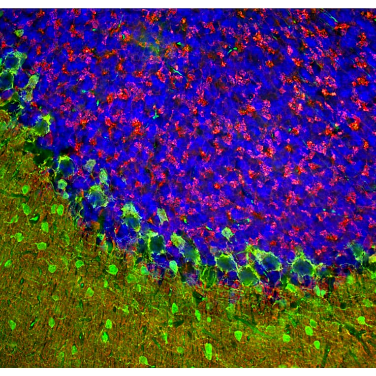
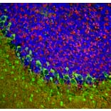
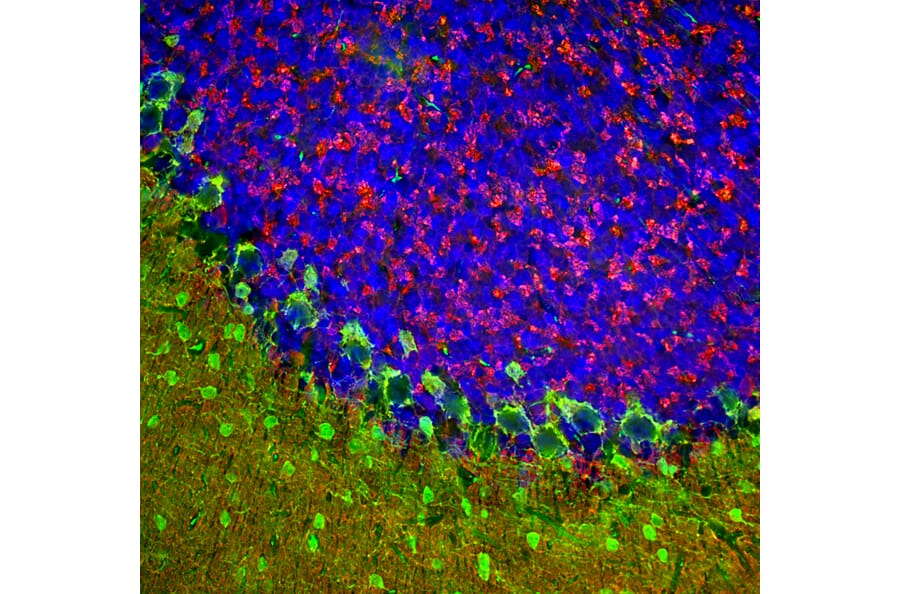
Immunofluorescent analysis of rat cerebellum section stained with Anti-beta Synuclein Antibody (A270581), at a dilution of 1:1,000, in red, and co-stained with Anti-Parvalbumin Antibody (A85316), at a dilution of 1:5,000, in green. Nuclear DNA is visualised in blue using Hoechst staining. Following transcardial perfusion with 4% paraformaldehyde, the brain was post-fixed for 24 hours, cut to 45 µm, and free-floating sections were stained using the above antibodies. Anti-beta Synuclein Antibody (A270581) detects protein concentrated in synaptic regions, and Anti-Parvalbumin Antibody (A85316) labels the perikarya and dendrites of Purkinje cells, and interneurons in the molecular layer of the cerebellum.
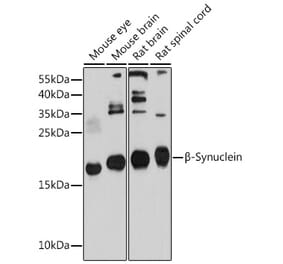
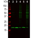
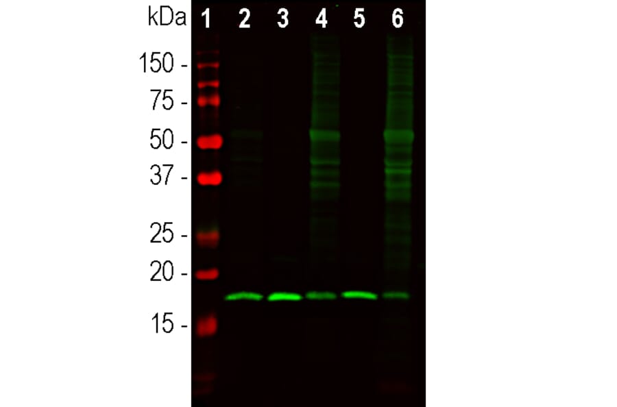
Western Blot - Anti-beta Synuclein Antibody (A270581)
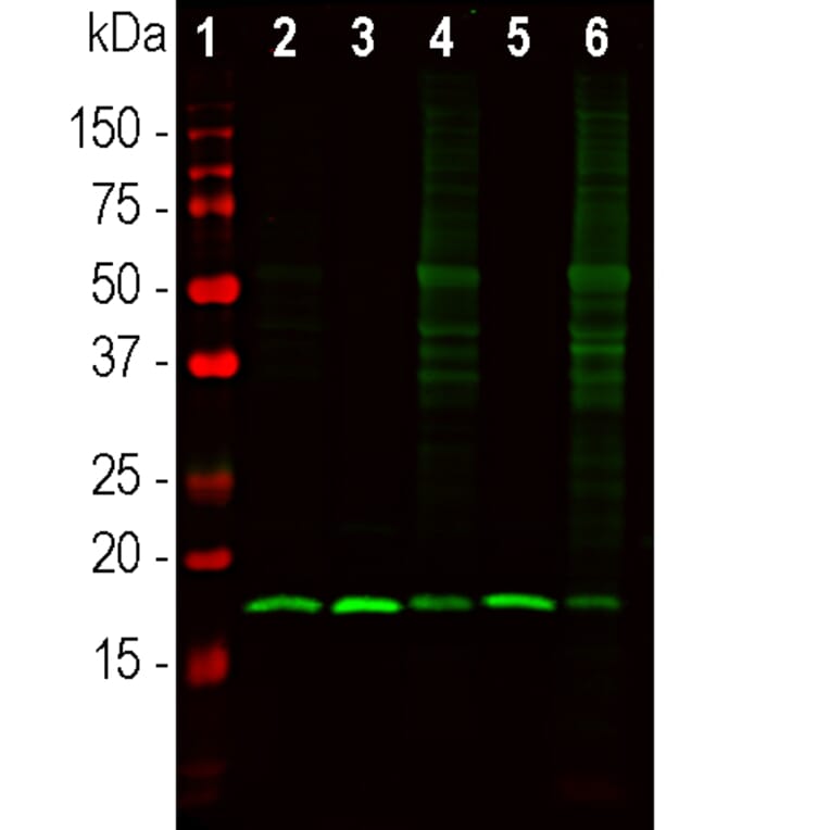
Western blot analysis of different tissue lysates using Anti-beta Synuclein Antibody (A270581), at a dilution of 1:1,000, in green. The lanes contain: [Lane 1] protein standard (red), [Lane 2] mouse cerebellum [Lane 3] mouse hippocampus, [Lane 4] rat cerebellum, [Lane 5] rat hippocampus, and [Lane 6] cow cerebellum. The strong band at about 17 kDa corresponds to the beta Synuclein protein.
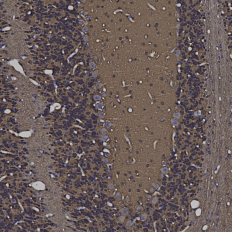
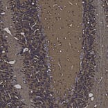
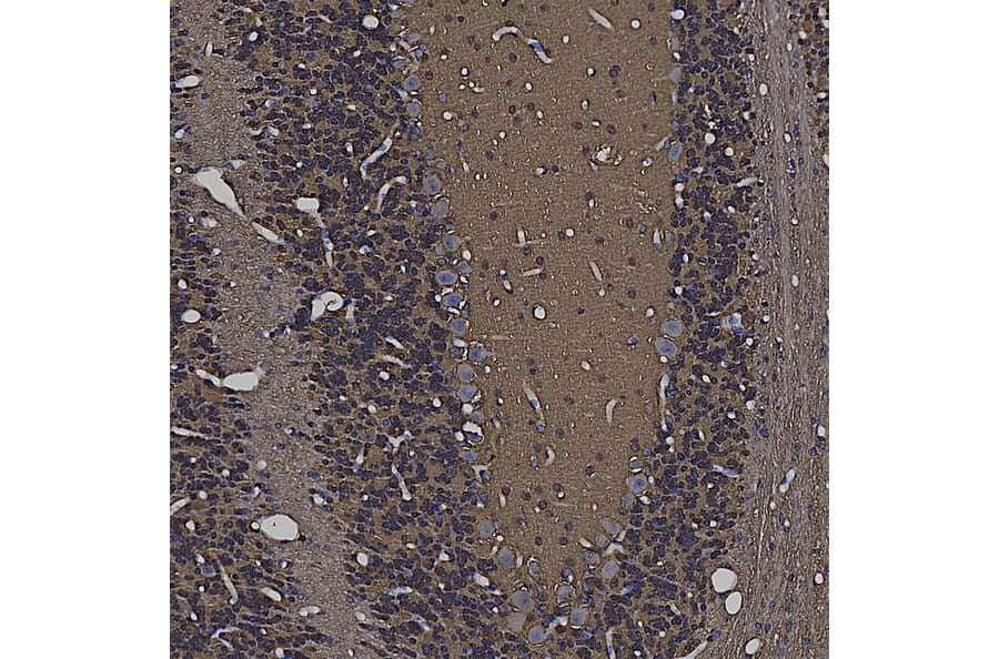
Immunohistochemistry analysis of a 4% PFA fixed paraffin embedded mouse hippocampus section with Anti-beta Synuclein Antibody (A270581) at a dilution of 1:2,000 detected with DAB (brown) using the Vector Labs ImmPRESS method and reagents with citra buffer retrieval. ß-synuclein is abundant in pre-synaptic regions of the brain. Note: this antibody performs well in testing with both 4% PFA and standard NBF fixed mouse, human, and rat tissues.
Publishing research using Anti-beta Synuclein Antibody (A270581)? Please let us know so that we can list the citation on this page.
Alternative products to Anti-beta Synuclein Antibody (A270581)


![Immunofluorescence - Anti-beta Synuclein Antibody [6A10] (A270558) - Antibodies.com](https://cdn.antibodies.com/image/catalog/270/A270558_1.jpg?profile=product_alternative)